Interferons-IFNs
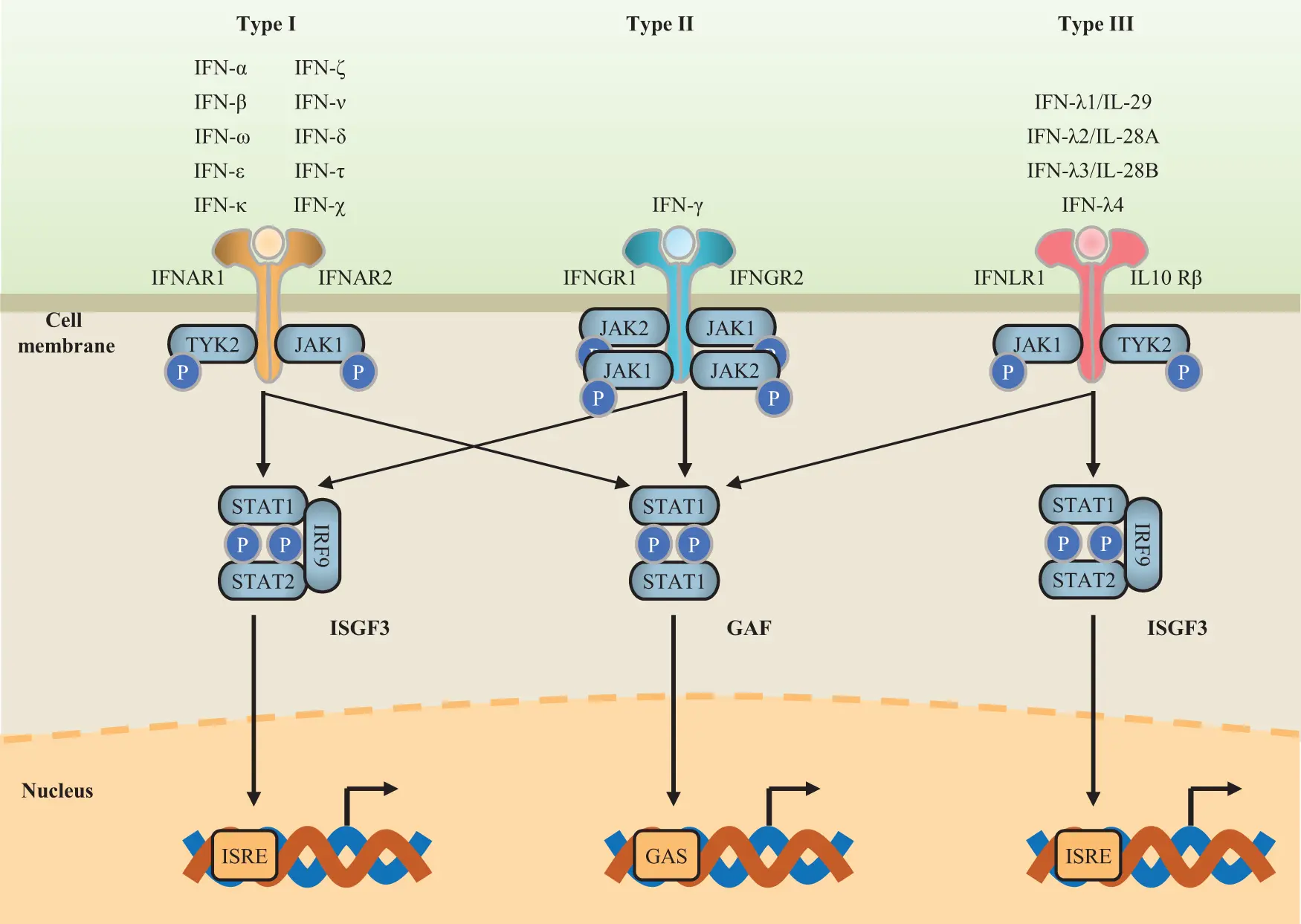
There are three families of IFNs: types I, II, and III. In humans, type I contains IFN-α (13 different subtypes), IFN-β (also known as IFN-β1), IFN-ε, IFN-κ, and IFN-ω.
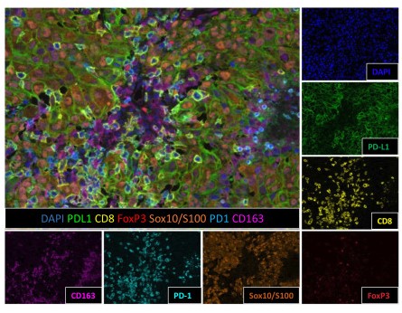
With the rapid development of tumor immunotherapy, precise assessment of the tumor microenvironment (TME) has become a core prerequisite for interpreting immune escape mechanisms, screening populations benefiting from immunotherapy, and optimizing treatment regimens. Traditional single-marker immunohistochemistry (IHC) and immunofluorescence (IF) techniques can only detect a single marker, failing to comprehensively present the complex composition of immune cell subsets, marker co-expression characteristics, and intercellular spatial relationships in the TME, which is insufficient to meet the needs of precise immunotherapy research and clinical applications. Against this backdrop, multiplex immunohistochemistry (mIHC) and multiplex immunofluorescence (mIF) technologies have emerged and rapidly iterated, becoming core technologies for parsing TME heterogeneity and mining immunotherapy biomarkers due to their advantage of simultaneously detecting multiple markers in tissue sections.
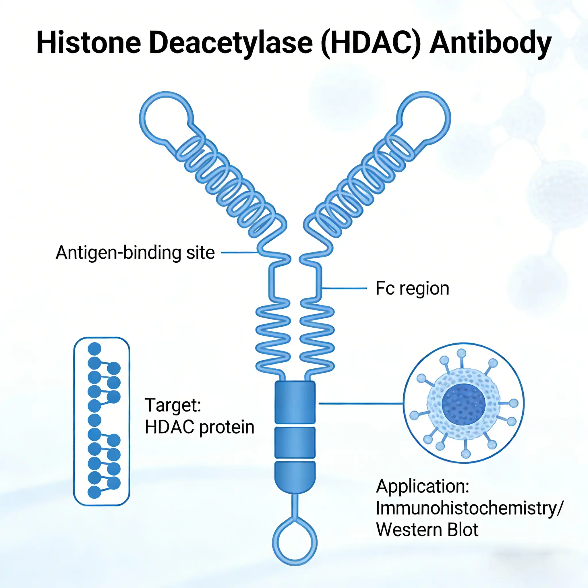
Acetylation of the histone tail causes chromatin to adopt an "open" conformation, allowing increased accessibility of transcription factors to DNA. The identification of histone acetyltransferases (HATs) and their large multiprotein complexes has yielded important insights into how these enzymes regulate transcription (1,2).