

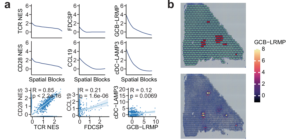
Literature Sharing: A Spatially Resolved Atlas of Gastric Cancer Reveals the Critical Role of Lymphocyte-Aggregated Regions
By integrating single-cell transcriptomics and spatial transcriptomics technologies, it constructs a spatially resolved cell atlas of gastric cancer, aiming to systematically reveal the spatial organization characteristics of the tumor microenvironment, the core role of tertiary lymphoid structures, and the spatial distribution patterns of immune checkpoint molecules. This provides a new perspective for the exploration of biomarkers for gastric cancer immunotherapy and a scientific basis for the formulation of clinical individualized immunotherapy strategies.
Literature Sharing: Multiplex Fluorescence Immunotechnology Reveals Colorectal Cancer Biomarkers
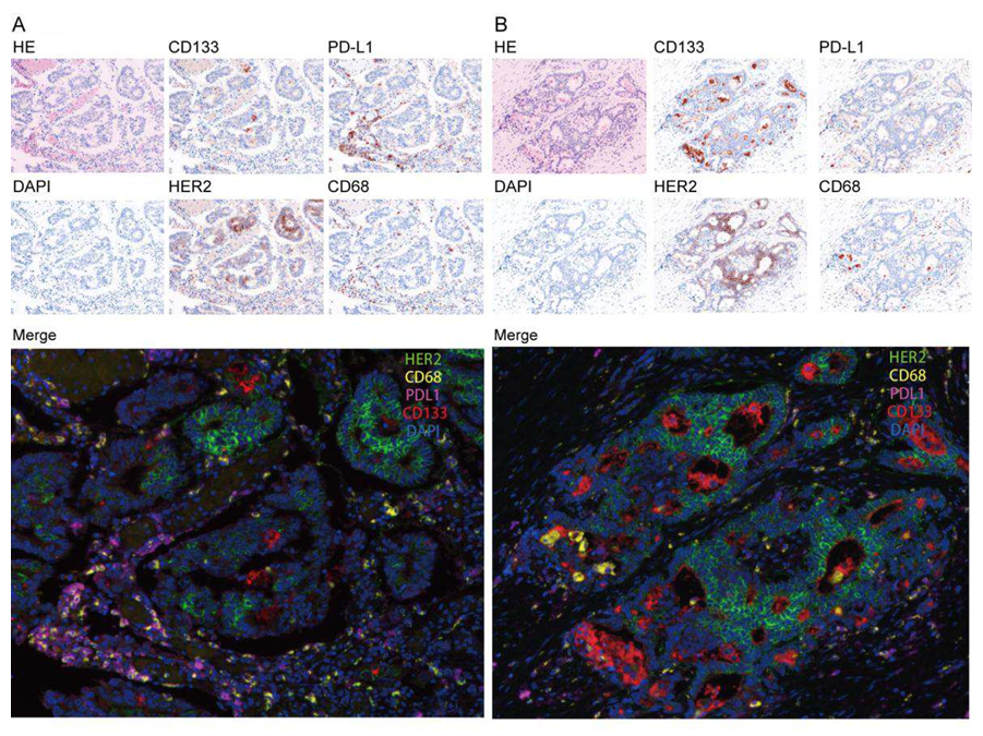
The authors innovatively used multiplex fluorescence immunohistochemistry (mIHC) technology, using tumor tissue and adjacent normal tissue from 84 colorectal cancer patients to construct tissue microarrays (TMA), simultaneously detecting the expression levels of cancer stem cell marker CD133, immune checkpoint molecule PD-L1, tumor-associated antigen HER2, and macrophage marker CD68, combining clinicopathological features with 7-year follow-up data to explore the diagnostic and prognostic value of these markers in colorectal cancer, providing new technical support and theoretical basis for precise diagnosis and treatment of colorectal cancer.
【ELISA Sample Processing Guide】Serum / Plasma
Serum and Plasma are samples obtained from blood through different processing methods. Due to the influence of the coagulation process, there are certain differences in their components, and these differences are reflected throughout sample preparation and storage. It is important to understand these before sample collection to choose the most appropriate sample preparation strategy.
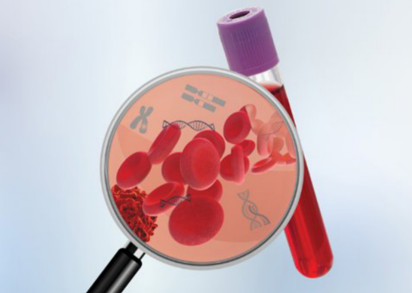
Reference Table for Serum and Plasma Enzyme Sources
The core principle of ELISA experiments is the specific binding of antigens and antibodies, while endogenous enzymes in samples can non-specifically catalyze substrate reactions or destroy antigen/antibody structures, leading to abnormal experimental signals and interference. Many customers often encounter issues such as biased test results, false positives or false negatives when conducting ELISA experiments, due to the failure to clarify the sources of various enzymes in the samples and add enzyme inhibitors in a targeted manner. To help customers accurately avoid this experimental pain point and efficiently complete ELISA experiments, EnkiLife has compiled the "Serum and Plasma Enzyme Source Reference Table", while combining core experimental needs to popularize the sources of serum and plasma enzymes and their association with ELISA experimental interference, helping everyone exclude interference and ensure reliable experimental results by clarifying enzyme sources and rationally adding enzyme inhibitors.
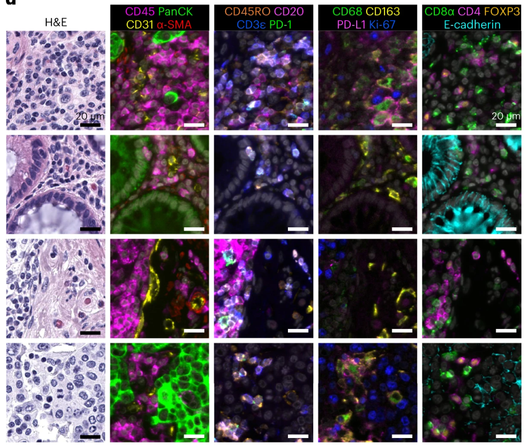
Literature Sharing: Combined Multiplex Immunofluorescence and Traditional Histology Imaging for Cancer Biomarker Discovery
Precise analysis of the tumor microenvironment is the core foundation for cancer diagnosis, prognosis assessment, and treatment decision-making. However, traditional tissue imaging techniques have obvious limitations: single immunofluorescence (IF) imaging can detect multiple molecular markers but lacks histological context, making it difficult to accurately localize cell types and distributions. Hematoxylin-Eosin (H&E) staining, as the gold standard for pathological diagnosis, clearly presents tissue morphology but cannot provide molecular-level immune characteristic information. Existing multiplex imaging techniques either have complex processes and are time-consuming, or have issues such as fluorescence crosstalk and insufficient reagent stability, making it difficult to achieve simultaneous precise detection of molecular markers + tissue morphology.

Loading Control Antibody
Housekeeping proteins perform numerous basic functions within the cell and are constitutively expressed at high levels in a variety of tissues and cell types. Western blot analysis commonly uses housekeeping proteins such as β-actin, COX IV, GAPDH, histone H3 and the α- and β-tubulins as loading controls.