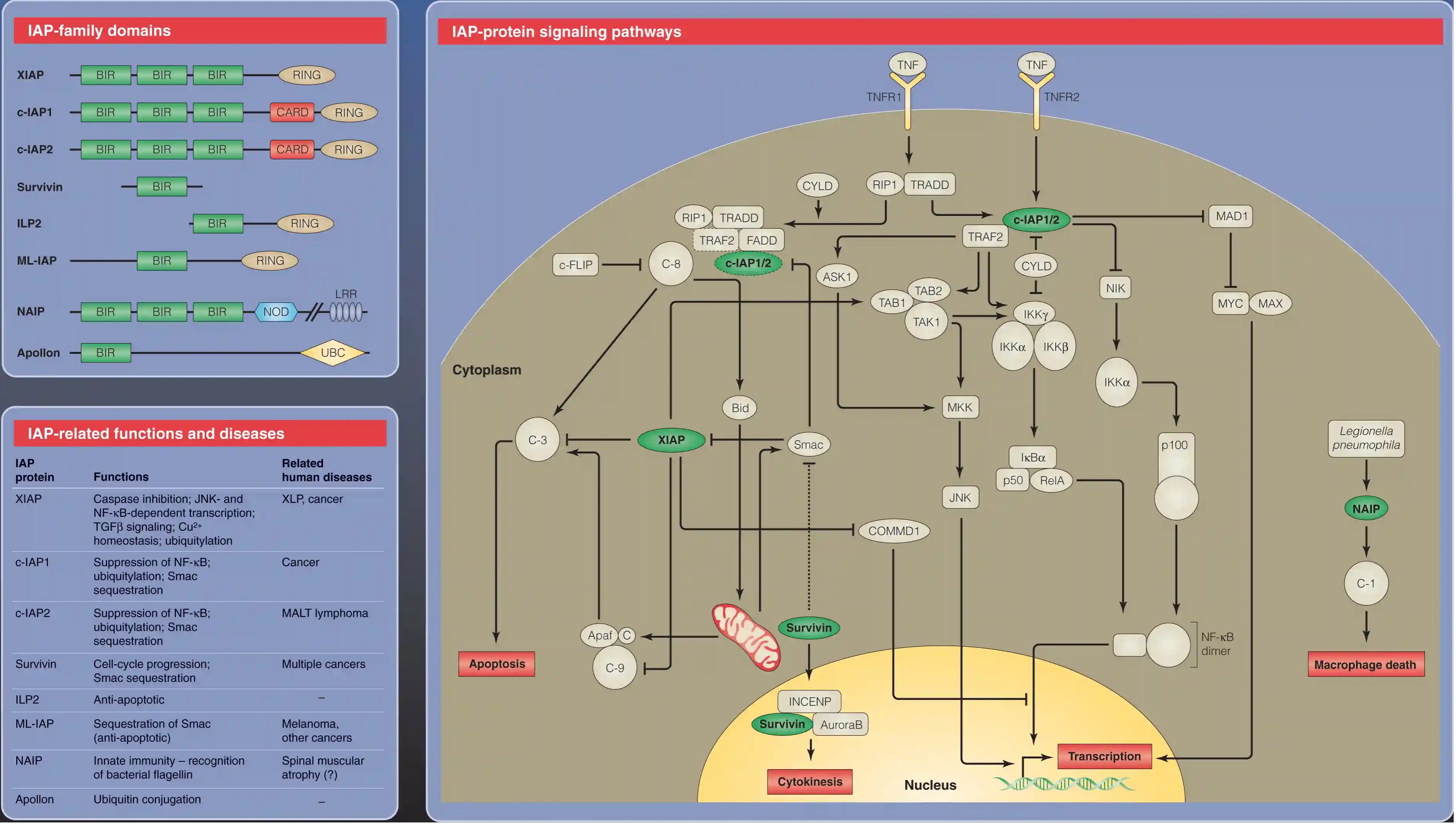
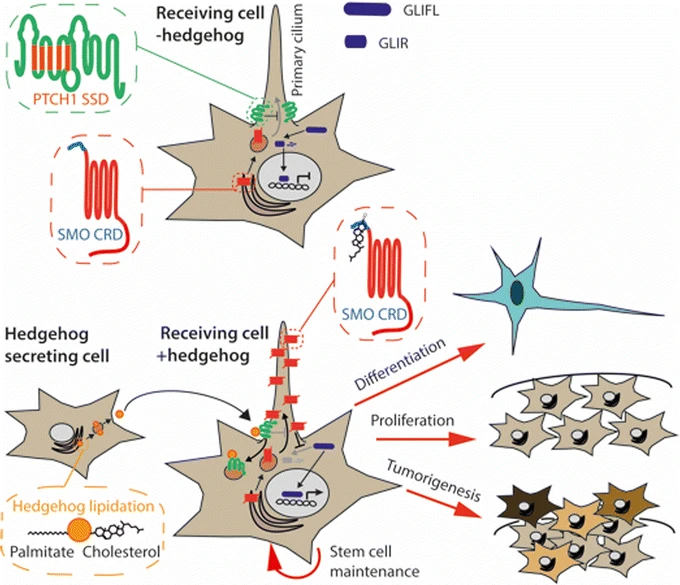
Hedgehog Signaling
Many of the genes that encode hedgehog pathway components have subsequently been associated with a range of inherited human developmental disorders and other pathologies.

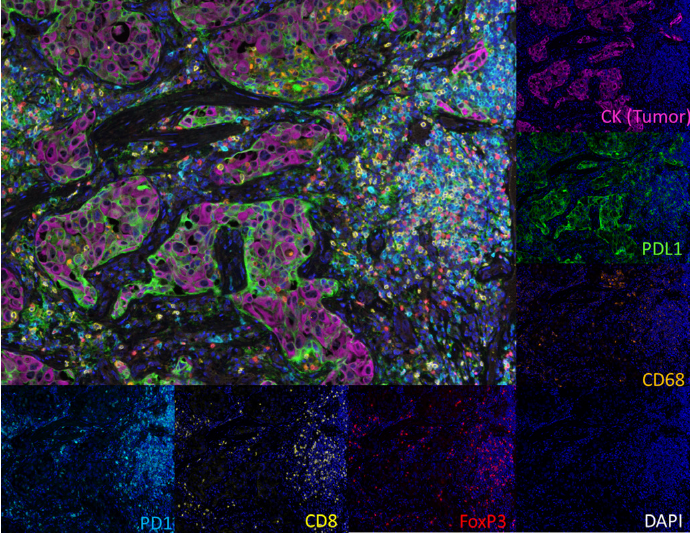
mIHC/mIF technology can quantify immune cell subsets, functional states, and spatial distribution, but its application has problems such as large platform differences and non-standardized operations. To this end, SITC convened experts from multiple fields to sort out the principles, processes, advantages and disadvantages of various mIHC/mIF technologies, formulate consensus recommendations for antibody selection, experimental optimization, validation and other links, and point out that this technology is expected to enter routine clinical applications, providing an important reference for its standardized development and biomarker research.
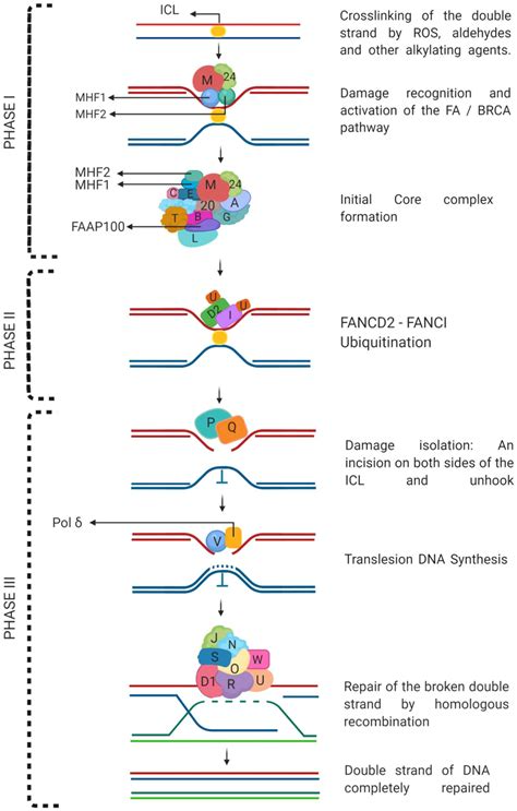
Fanconi anemia (FA) is an autosomal recessive genetic disorder resulting in symptoms that include chromosomal breakage, bone marrow failure, hypersensitivity to DNA cross-linking agents (such as mitomycin C), and a predisposition to cancer (1). In response to DNA damage, the FA nuclear complex (FANCA, FANCB, FANCC, FANCE, FANCF, FANCG, FANCM) induces mono-ubiquitination of FANCD2 and FANCI (2). Monoubiquitination of FANCD2 induces localization of FANCD2 to sites of DNA damage, where it interacts with BRCA1. FANCJ/BRIP1, FANCD1/BRCA2, and FANCN/PALB2 are also recruited to sites of DNA damage (3).
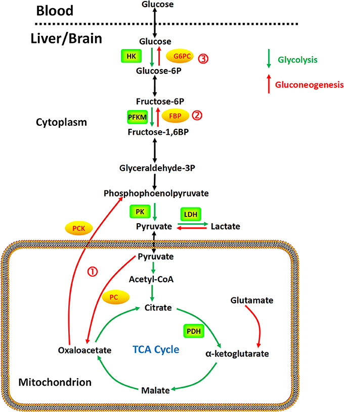
This document focuses on gluconeogenesis and its associated key enzymes, along with relevant antibodies and research references. It elaborates on the functions of multiple enzymes involved in gluconeogenesis and glycolysis, such as enolase isoforms (ENO1/2/3), fructose-1,6-bisphosphatase 1 (FBP1), glucose-6-phosphate isomerase (GPI), phosphoenolpyruvate carboxykinase (PCK1/2), phosphoglycerate mutase 1 (PGAM1), phosphoglycerate kinase 1 (PGK1), and pyruvate carboxylase (PC). These enzymes play pivotal roles in metabolic processes, with abnormal expression linked to tumor progression (e.g., breast cancer, lung cancer, clear cell renal cell carcinoma) and other diseases. Additionally, the document presents a regulatory diagram of glucose metabolism (encompassing glycolysis, gluconeogenesis, and the tricarboxylic acid cycle) and provides detailed information on specific antibodies targeting the aforementioned enzymes, including their catalog numbers, reactivities, and applications. Relevant research references are also included to support the discussed findings.
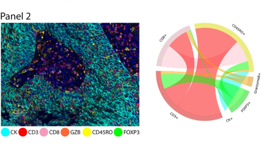
Although tumor immunotherapy has developed rapidly and achieved significant results, malignant tumors can escape immune surveillance by constructing an immunosuppressive microenvironment, which severely limits treatment efficacy. After the discovery of immune checkpoint molecules such as PD-1 and CTLA-4 and their roles in tumor immune evasion, clarifying the interaction between immune cells and cancer cells has become a core prerequisite for developing novel immunotherapies. Multiplex immunofluorescence (mIF) is a reliable high-throughput method that can directly observe multiple biomarkers expressed by individual cells and analyze the spatial relationships of these biomarkers in different cell populations, which is impossible with traditional immunohistochemistry (IHC) techniques. Therefore, the study "Immuno-profiling and cellular spatial analysis using five immune oncology multiplex immunofluorescence panels for paraffin tumor tissue" can identify multiple cell subpopulations by combining carefully selected antibodies. The tyramide signal amplification (TSA) manual protocol was used as a standard reference for validating multiplex staining. The automated stainer significantly reduced the original 4-5 day manual staining time to 14-17 hours while improving staining consistency. The literature demonstrates the optimization process and reproducibility of automated TSA staining, and validates its application value in tumor microenvironment research and cellular phenotype spatial distribution analysis in a small cohort of non-small cell lung cancer (NSCLC) samples.