
Nearly all research on protein function, expression, localization, modification and interaction starts with a critical step: releasing proteins from complex cellular or tissue environments, namely cell lysis and protein extraction. A successful lysis protocol must achieve two seemingly contradictory goals: first, it must be sufficiently harsh to thoroughly break cell membranes, cell walls (for plants and certain microorganisms) and tissue matrix barriers; second, it must be gentle enough to maximally protect target proteins from degradation, denaturation, oxidation or non-specific aggregation, so as to retain their native structure and biological activity.
However, the diversity of biological samples poses great challenges to this process. Tough plant cell walls, animal muscle rich in connective tissue, blood samples filled with highly abundant interfering proteins, and fragile cultured cells all feature distinct physical and chemical properties, which means there is no universal lysis protocol. Improper protocols may lead to low protein yield, incomplete extraction of specific proteins (such as membrane proteins and nuclear proteins), degradation of proteins by endogenous proteases, or lysis products incompatible with downstream analytical techniques.
Cell lysis and protein extraction are key procedures for cellular protein research, laying a foundation for numerous downstream applications including western blotting, enzyme activity assays and mass spectrometry.
1. Low temperature throughout all operations: Perform all procedures on ice and pre-cool lysis buffer to prevent protein degradation.
2. Timely addition of inhibitors: Freshly add protease inhibitors (PMSF, protease inhibitor cocktail) right before use. Phosphatase inhibitors (sodium vanadate, sodium fluoride, β-glycerophosphate) are required for phosphorylated protein research.
3. Auxiliary mechanical disruption: Tissue samples cannot release sufficient proteins with lysis buffer alone; physical methods such as grinding, sonication and homogenization are required.
4. Lysis duration control: Lyse on ice for 30~60 min; excessively long incubation will cause protein degradation.
5. Clarification by centrifugation: Centrifuge at 12000~14000 g for 10~15 min at 4°C after lysis; the supernatant is total protein solution.
- Complete extraction of macromolecular proteins.
- Preserve post-translational protein modifications including phosphorylation, glycosylation, ubiquitination, methylation and acetylation.
- One-step protocol without extra enzyme inhibitors.
- Compatible with both cell and tissue samples.
Validation Data: Sample Compatibility Assay
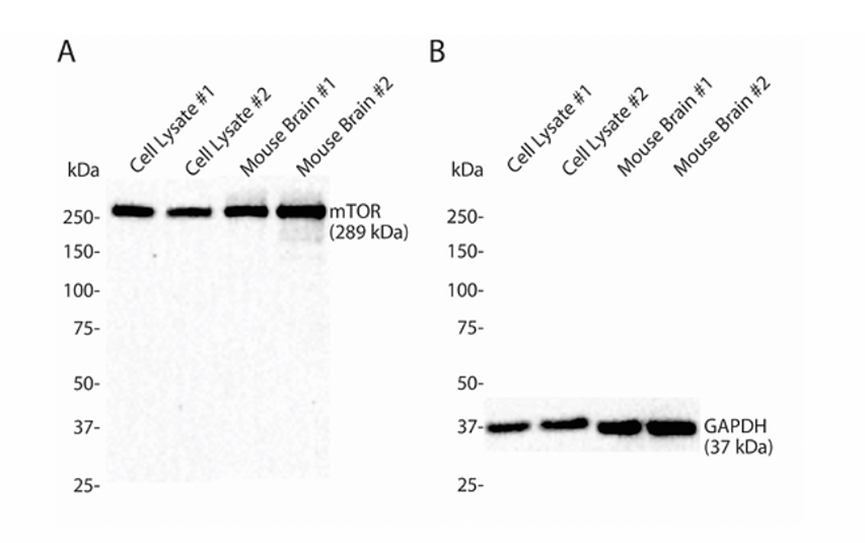
Figure A: Western blot with mTOR (289 kDa) antibody on lysates of HeLa cells and mouse brain tissue (2 replicates each) prepared with RC0005; loading quantity: 50 μg lysate per lane.
Figure B: Western blot with GAPDH (37 kDa) antibody on lysates of HeLa cells and mouse brain tissue (2 replicates each) prepared with RC0005; loading quantity: 50 μg lysate per lane.
Data demonstrates that RC0005 delivers excellent lysis performance for both cell and tissue samples, compatible with proteins of various molecular weights.
Cell samples feature simple structures without extracellular matrix barriers, thus requiring the least rigorous lysis. They are divided into adherent cells and suspension cells.
| Sample Treatment | Discard culture medium; wash 2-3 times with PBS |
|---|---|
| Recommended Lysis Buffer | RIPA (strong, complete lysis) or NP-40 (mild, preserve protein complexes) |
| Lysis Method | Incubate on ice for 30 min |
| Centrifugation Parameters | 12,000-14,000 rpm, 4°C, 15 min; collect supernatant |
| Tips | Lysis can be performed directly in culture dishes; scrape cells after adding lysis buffer |
| Sample Treatment | Harvest cells by centrifugation (500-1000 rpm, 5 min); wash 1-2 times with PBS |
|---|---|
| Recommended Lysis Buffer | NP-40 or RIPA |
| Lysis Method | Incubate on ice for 30 min; pipette up and down thoroughly every 10 min to ensure full cell lysis |
| Centrifugation Parameters | 12,000 rpm, 4°C, 10-15 min |
Animal tissues contain abundant extracellular matrix with varying compactness. Disruption intensity and buffer strength shall be selected according to tissue characteristics. Core rule: denser and more fibrous tissues require stronger mechanical disruption; tissues with high endogenous enzyme activity need sufficient inhibitors.
| Sample Characteristics | Extremely high lipid content (abundant myelin sheath), moderate protein abundance, medium endogenous protease activity, soft texture |
|---|---|
| Sample Treatment | Place freshly dissected tissue on ice immediately; remove meninges and blood vessels; separate subregions (cortex, hippocampus, cerebellum, etc.) if needed |
| Recommended Lysis Buffer | Strong RIPA supplemented with 1% SDS (enhanced membrane protein solubilization) |
| Mechanical Disruption | Homogenize tissue until no visible solid fragments remain |
| Lysis Method | Incubate on ice for 30 min, vortex intermittently 2-3 times |
| Centrifugation Parameters | 12,000-14,000 rpm, 4°C, 15-20 min; collect intermediate clear layer, avoid upper lipid layer and lower pellet |
| Tips | A white lipid layer forms on top after centrifugation due to high brain lipid content; avoid aspirating lipid to prevent interference with subsequent WB experiments. |
| Sample Characteristics | Extremely high protein content, potent endogenous proteases and phosphatases, soft texture easy to disrupt |
|---|---|
| Sample Treatment | Remove gallbladder and connective tissue; flush with PBS to eliminate blood, blot dry with filter paper |
| Recommended Lysis Buffer | Standard RIPA with double dosage of protease and phosphatase inhibitors |
| Mechanical Disruption | Homogenize tissue until no visible solid fragments remain |
| Lysis Method | Lyse on ice for 30 min, vortex intermittently 2-3 times |
| Centrifugation Parameters | 12,000-14,000 rpm, 4°C, 15 min; collect supernatant |
| Tips | · Hepatic protease activity is extremely high; add PMSF immediately before use with concentration elevated to 2 mM. · Blood interferes with protein quantification; thoroughly perfuse or wash tissue with PBS. · Liver protein concentration is high; increase dilution factor for subsequent quantification. |
| Sample Characteristics | Abundant muscle fibers, dense tissue with high mechanical resistance; regular homogenization insufficient for full disruption; rich mitochondrial proteins |
|---|---|
| Recommended Lysis Buffer | Strong RIPA with auxiliary sonication |
| Mechanical Disruption | Liquid nitrogen grinding is optimal; grind until no particulate matter exists |
| Lysis Method | Lyse on ice for 30 min with intermittent sonication assistance |
| Centrifugation Parameters | 12,000-14,000 rpm, 4°C, 20 min |
| Tips | · Liquid nitrogen grinding is the most effective method for muscle tissue; incomplete grinding results in extremely low protein yield. · Avoid foam generation during sonication to prevent protein oxidation and denaturation. |
| Sample Characteristics | Dense vascular network with massive residual blood; distinct protein profiles between cortex (glomeruli, renal tubules) and medulla; moderately tough tissue with medium extracellular matrix content; strong endogenous protease and phosphatase activity, abundant membrane transporters, ion channels and receptors |
|---|---|
| Sample Treatment | Remove renal capsule; separate cortex and medulla for targeted analysis |
| Recommended Lysis Buffer | Strong RIPA; enhanced RIPA with 1% SDS for membrane transporter research |
| Mechanical Disruption | Homogenize tissue until no visible solid fragments; tough samples may be ground into fine powder in liquid nitrogen before buffer resuspension |
| Lysis Method | Lyse on ice for 30 min, vortex thoroughly every 10 min |
| Centrifugation Parameters | 12,000 rpm, 4°C, 15-20 min; aspirate intermediate clear supernatant, discard bottom debris pellet and surface lipid film |
| Tips | · Residual blood is the primary interference factor for kidney samples; in vivo perfusion or repeated ex vivo rinsing with PBS is required to eliminate hemoglobin and albumin masking endogenous tissue proteins. · Cortex and medulla exhibit divergent protein expression; renal cortex is preferred for general total protein extraction for higher target protein abundance and stable results. · High endogenous phosphatase activity requires freshly prepared sufficient phosphatase inhibitors and strict low-temperature operation to avoid loss of phosphorylation signals. |
| Sample Characteristics | Extremely high lipid proportion with minimal protein content; standard protocols yield low protein recovery |
|---|---|
| Sample Treatment | Remove blood vessels and connective tissue; trim excess fat; rinse with PBS and mince finely |
| Recommended Lysis Buffer | Lysis buffer containing 1-2% SDS (high detergent concentration required for lipid solubilization) |
| Mechanical Disruption | Homogenize tissue until no visible solid fragments remain |
| Lysis Method | Lyse on ice for 30 min |
| Centrifugation Parameters | 12,000 rpm, 4°C, 20 min; carefully aspirate lower aqueous protein phase, fully discard upper lipid layer. Chloroform-methanol precipitation may be applied to remove residual lipids if needed. |
| Tips | · Larger initial sample mass is required due to low protein yield of adipose tissue. · Distinct separation between lipid and aqueous phases post-centrifugation; avoid aspirating lipid with pipette tip. · Residual lipid causes smearing and tailing bands in SDS-PAGE electrophoresis. |
| Sample Characteristics | Highly developed sinus system with massive residual blood and abundant erythrocytes; soft tissue with potent endogenous proteases and nucleases, prone to protein degradation; blood proteins easily mask intrinsic tissue protein signals |
|---|---|
| Sample Treatment | Rinse spleen repeatedly with pre-cooled PBS, gently squeeze to drain residual sinus blood; cardiac perfusion is preferred for complete blood elimination if available |
| Recommended Lysis Buffer | Strong RIPA lysis buffer |
| Mechanical Disruption | Homogenize tissue until no visible solid fragments remain |
| Lysis Method | Static lysis on ice for 30 min |
| Centrifugation Parameters | 12,000 rpm, 4°C, 15-20 min; aspirate intermediate clear protein supernatant, discard bottom cell and erythrocyte pellet |
| Tips | · Strictly control blood contamination: cardiac perfusion is optimal; perform secondary low-speed centrifugation if supernatant remains reddish to remove residual erythrocytes. · Prevent protein degradation: high protease activity in immune cells; snap-freeze in liquid nitrogen immediately after dissection or maintain full ice incubation without room-temperature exposure. |
| Sample Characteristics | (1) Distinct protein profiles across tissue layers; (2) High risk of contamination from luminal debris; (3) Extremely high risk of protein degradation |
|---|---|
| Sample Treatment | Dissect corresponding intestinal segments (duodenum, jejunum, ileum, colon) as required by experiment; cut longitudinally and remove luminal contents; rinse gently with PBS; separate mucosal and muscular layers if needed |
| Recommended Lysis Buffer | RIPA lysis buffer |
| Mechanical Disruption | (1) Mucosa samples: homogenize until no visible solid fragments. (2) Full-thickness / muscular samples: grind into fine powder in liquid nitrogen before buffer resuspension |
| Lysis Conditions | Static lysis on ice for 30 min, vortex gently every 10 min |
| Centrifugation Parameters | 12,000 rpm, 4°C, 15 min; repeat centrifugation or brief sonication if supernatant remains turbid and viscous |
| Tips | · Thorough rinsing is required to eliminate mucus interference. · Diverse and highly active intestinal proteases necessitate broad-spectrum protease inhibitor cocktail; elevate PMSF final concentration to 2 mM. |
| Sample Type | Characteristics | Processing Protocol |
|---|---|---|
| Whole Blood | Contains erythrocytes, leukocytes, platelets and plasma proteins |
|
| Serum | Supernatant depleted of blood cells and fibrinogen | Incubate fresh whole blood at room temperature for 30~60 min for complete coagulation; centrifuge at 3000 g, 4°C for 15 min; carefully aspirate pale yellow clear upper layer, avoid touching intermediate leukocyte layer and bottom erythrocyte pellet. Direct dilution for use without additional lysis. |
| Plasma | Contains anticoagulant with preserved fibrinogen | Invert EDTA-anticoagulated whole blood for uniform mixing; centrifuge at 3000 g, 4°C for 15 min; collect upper plasma layer. Direct dilution for use without additional lysis. |
| Blood Cells (PBMC / Leukocytes) | Requires erythrocyte lysis prior to isolation | 1. PBMC Isolation via Density Gradient Centrifugation (1) Dilute fresh EDTA whole blood with equal volume PBS, slowly layer above Ficoll-Paque separation liquid without disrupting the interface. (2) Centrifuge at 2000 g, room temperature for 20 min with minimal acceleration/deceleration. (3) Gently aspirate intermediate milky PBMC buffy coat, transfer to new tube and resuspend in 5-10 volumes pre-cooled PBS for washing. (4) Centrifuge at 1500 g, 4°C for 10 min, discard supernatant; repeat washing 1-2 times to fully remove residual platelets and separation medium. 2. Cell Lysis: Add 100~200 μL pre-cooled lysis buffer per 1×10⁷ cells, pipette to homogenize, lyse on ice for 30 min with gentle vortex every 10 min. 3. Clarification Centrifugation: Centrifuge at 14000 g, 4°C for 15 min, aspirate intermediate clear protein supernatant and discard bottom cell debris pellet. |
Plant samples feature rigid cell walls and abundant polyphenols, polysaccharides and secondary metabolites, which easily induce protein browning and precipitation, making them the most difficult sample type for lysis. Core strategy: liquid nitrogen grinding for cell wall disruption plus polyphenol/polysaccharide removal measures. Plant proteins oxidize rapidly; utilize extracted protein within 1 month post-lysis. Unlysed samples: store in liquid nitrogen if available, -80°C as secondary option, and avoid storage longer than 1 month at -20°C as last resort.
| Step | Operation | Critical Notes |
|---|---|---|
| Mortar Pre-cooling | Pre-chill mortar with liquid nitrogen | Dry grinding is forbidden; replenish liquid nitrogen when depleted |
| Sample Collection | Tissue preserved at -80°C or liquid nitrogen | Freeze immediately in liquid nitrogen post-dissection |
| Grinding | Grind tissue into fine powder under liquid nitrogen | Maintain low temperature to prevent tissue thawing |
| Powder Collection | Transfer powder with pre-cooled spatula after nitrogen evaporation | Transfer powder into centrifuge tube supplemented with β-mercaptoethanol |
| Lysis | Add lysis buffer at 99:1 ratio plus sonication disruption | No additional homogenization required |
| Incubation | Incubate at 4°C for 20-30 min | Allow sufficient protein release |
| Centrifugation | Centrifuge 12,000-14,000 rpm for 15-20 min, collect supernatant | White pellet at bottom may be frozen for backup |
| Sample Characteristics | Thin cell walls easy to grind; abundant chloroplasts with highly enriched Rubisco masking low-abundance proteins; moderate polyphenol content |
|---|---|
| Sample Treatment | Snap-freeze in liquid nitrogen and grind into fine powder |
| Recommended Lysis Buffer | Plant RIPA supplemented with 1% PVP and 10 mM β-mercaptoethanol |
| Mechanical Disruption | Liquid nitrogen grinding is preferred |
| Centrifugation Parameters | 12,000-14,000 rpm, 4°C, 15-20 min, collect supernatant; repeat centrifugation if supernatant retains pigmentation |
| Tips | · Commercial plant protein enrichment kits may be used to deplete abundant Rubisco and improve detection sensitivity of low-abundance proteins. · Young leaves yield intact proteins more readily than senescent leaves. · Chlorophyll interferes with BCA quantification; Bradford or 2-D quantification kits are recommended. |
| Sample Characteristics | Fibrous tissue with high impurity load, elevated polysaccharide and secondary metabolite levels prone to browning; soil contamination on root surfaces |
|---|---|
| Sample Treatment | Thoroughly rinse soil residue, snap-freeze in liquid nitrogen and grind to fine powder |
| Recommended Lysis Buffer | Buffer containing 2% PVP and 5% glycerol, paired with TCA-acetone purification precipitation |
| Mechanical Disruption | Liquid nitrogen grinding |
| Centrifugation Parameters | 12,000 rpm, 4°C, 15 min |
| Tips | · Direct lysis yields high impurity levels impairing electrophoresis quality; TCA-acetone precipitation purification is recommended. · Young root tips contain higher protein concentrations with less lignification, prioritize these segments. |
| Sample Characteristics | High lignification with abundant rigid fibers and thick cell walls difficult to grind; lower total protein content compared to leaves |
|---|---|
| Sample Treatment | Remove epidermis, cut young segments into small pieces, snap-freeze in liquid nitrogen |
| Recommended Lysis Buffer | Strong lysis formulation supplemented with 2% SDS |
| Mechanical Disruption | Liquid nitrogen grinding (mandatory for lignified tissue) |
| Centrifugation Parameters | 14,000 rpm, 4°C, 20 min |
| Tips | · Fully grind lignified stems until no fibrous particles are visible to naked eye. · Heat-assisted lysis significantly improves protein yield but is incompatible with experiments requiring native protein activity retention. |
| Sample Characteristics | Flowers exhibit the highest structural heterogeneity and severe secondary metabolite interference among plant reproductive organs; primary lysis challenges include browning suppression and low-abundance protein enrichment |
|---|---|
| Sample Treatment | Place freshly harvested flowers on ice immediately; isolate target compartments (petals, stamens, pistils) as experimental requirements dictate, discard wilted tissue and surface contaminants. Rinse briefly with pre-cooled distilled water, blot moisture with filter paper, minimize room-temperature exposure duration. |
| Mechanical Disruption | Liquid nitrogen grinding |
| Lysis Method 1: Direct Lysis | Suitable for lightly browning tissues such as stamens and receptacles: · Add pre-cooled lysis buffer at mass:volume ratio of 1:5~1:8, vortex rapidly and transfer to ice instantly. · Lyse on ice for 30 min, vortex gently every 10 min. · Centrifuge at 14000 g, 4°C for 20 min, aspirate intermediate clear protein supernatant, discard bottom cell debris and surface pigment flotation layer. · Add small amounts of PVPP to adsorb polyphenols and re-centrifuge if supernatant retains prominent pigmentation. |
| Lysis Method 2: TCA-Acetone Precipitation | Preferred for high-pigment tissues including petals: · Transfer ground powder to pre-cooled EP tube, add 3 volumes pre-cooled 10% TCA-acetone solution supplemented with 0.07% β-mercaptoethanol, mix thoroughly. · Precipitate at -20°C for minimum 2 h (or overnight), invert to homogenize 1~2 times during incubation. · Centrifuge at 12000 g, 4°C for 15 min and discard supernatant. · Wash pellet 3 times with pre-cooled 80% acetone, fully discard supernatant each cycle to eliminate residual TCA. · Air-dry under ventilation for 5~10 min until acetone odor dissipates (avoid over-drying which impairs protein resolubilization). · Resuspend pellet in lysis buffer, incubate on ice for 30 min to facilitate solubilization. · Centrifuge at 14000 g for 15 min, collect supernatant as purified protein sample. |
| Sample Characteristics | Primary challenges include hard seed coat disruption, removal of abundant storage contaminants, and protein degradation induced by endogenous enzyme activation |
|---|---|
| Sample Treatment | Remove seed coat and isolate embryo/endosperm fractions per experimental design; rinse surface dust rapidly with distilled water and blot dry. Grind dry seeds directly in liquid nitrogen; hydration soaking is forbidden to prevent endogenous enzyme-mediated protein degradation. |
| Recommended Lysis Buffer | 1. Starch/Protein Seeds: Strong lysis buffer with 2% SDS or plant strong RIPA; TCA-acetone precipitation for high-purity requirements. 2. Oilseeds: Strong RIPA with mandatory n-hexane / petroleum ether degreasing pre-treatment |
| Mechanical Disruption | Liquid nitrogen grinding |
| Lysis Method 1: Starch/Protein Seeds | Rice, wheat, beans, etc.: · Add pre-cooled lysis buffer at mass:volume ratio of 1:10, vortex rapidly and lyse on ice for 30 min with vortexing every 10 min. · Centrifuge at 14000 g, 4°C for 20 min, aspirate intermediate clear supernatant; repeat centrifugation if heavy starch pellet accumulates at bottom. · TCA-acetone precipitation (identical to flower protocol) directly post-grinding for high-purity experiments to fully eliminate starch, polysaccharide and polyphenol contaminants. |
| Lysis Method 2: Oilseeds | Peanuts, rapeseed, sesame, etc.: · Mix ground powder with pre-cooled n-hexane, vortex and incubate 10 min; centrifuge at 12000 g for 5 min and discard supernatant; repeat degreasing 2~3 cycles. · Air-dry under ventilation for ~5 min to eliminate organic solvent residues and avoid protein denaturation. · Add pre-cooled strong RIPA, vortex and lyse on ice for 30 min. · Centrifuge at 14000 g, 4°C for 20 min, carefully aspirate lower aqueous protein phase and fully discard surface residual lipid layer. |
1. Inhibitors must be freshly supplemented: PMSF rapidly degrades in aqueous solution; add to RIPA immediately prior to use, pre-mixed storage is prohibited.
3. Mandatory low-temperature operation: Perform all steps on ice or at 4°C; heat generation during RIPA lysis induces protein degradation and loss of phosphorylation sites.
4. Avoid excessive sample loading: Overabundant tissue/cells relative to insufficient lysis buffer causes incomplete lysis, low protein concentration and high impurity background.
5. Standard supernatant collection protocol: Only aspirate fully clarified supernatant post-centrifugation; strictly avoid lipid layers and pellets to prevent non-specific bands and high background in downstream WB.
| Catalog Number | Product Name |
|---|---|
| RA10020 | 2-Hour Rapid Western Blot Ready-to-Use Complete Workflow Kit |
| RA10042 | Streamlined Western Blot Complete Kit |
| G01420 | Bis-Tris,4-20% |
| RA10043 | RapidSet Broad-Range Gradient Gel Kit |
| RC0005 | Cell/Tissue Lysis Buffer |
| RC0160 | RIPA Lysis Buffer (strong) |
| RC0161 | RIPA Lysis Buffer (medium) |
| RC0162 | RIPA Lysis Buffer (weak) |
| RA10063 | Protease & Phosphatase Inhibitor Cocktail (100×, EDTA-free) |
| RA10066 | Protease Inhibitor Cocktail (100×, EDTA-free) |
| RA10067 | Phosphatase Inhibitor Cocktail II (50×) |
| BC00006 | BCA Protein Assay Kit (BCA Method) |
| RA10055 | 5×Loading Buffer |
| RA10037 | Calibrated Color Prestained Protein Marker (8-180kDa) |
| RA10038 | Calibrated Color Prestained Protein Marker (10-250kDa) |
| RA10039 | Calibrated High Molecular Weight Color Prestained Protein Marker (25–400 kDa) |
| RA10044 | 20× Universal Rapid Electrophoresis Buffer |
| RA10051 | 20× Ice-Free Rapid Transfer Buffer |
| RA022 | PVDF Membrane (0.22μm) |
| RA045 | PVDF Membrane(0.45μm) |
| RA10052 | 5× Protein-Free Rapid Blocking Diluent |
| RA10058 | 5min Protein-Free Rapid Blocking Solution |
| RA10059 | 1min Protein-Free Rapid Blocking Solution |
| RA10056 | No-Blocking Rapid Antibody Diluent |
| RA10001 | Super-sensitive ECL chemiluminescent reagent |
| RA10041 | Antibody Stripping Buffer |
| RA10057 | Coomassie Brilliant Blue Fast Staining Solution (Non-Dehydration) |
| RA10061 | 10×TBST |