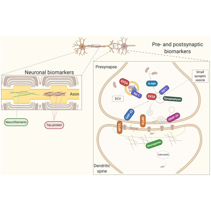
Pre-synaptic and Post-synaptic Markers
Synapses are the site for brain communication where information is transmitted between neurons and stored for memory formation.
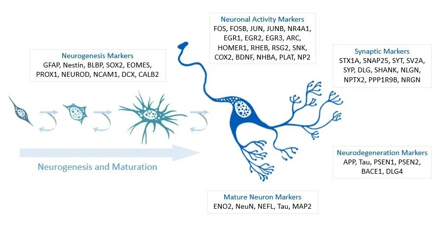
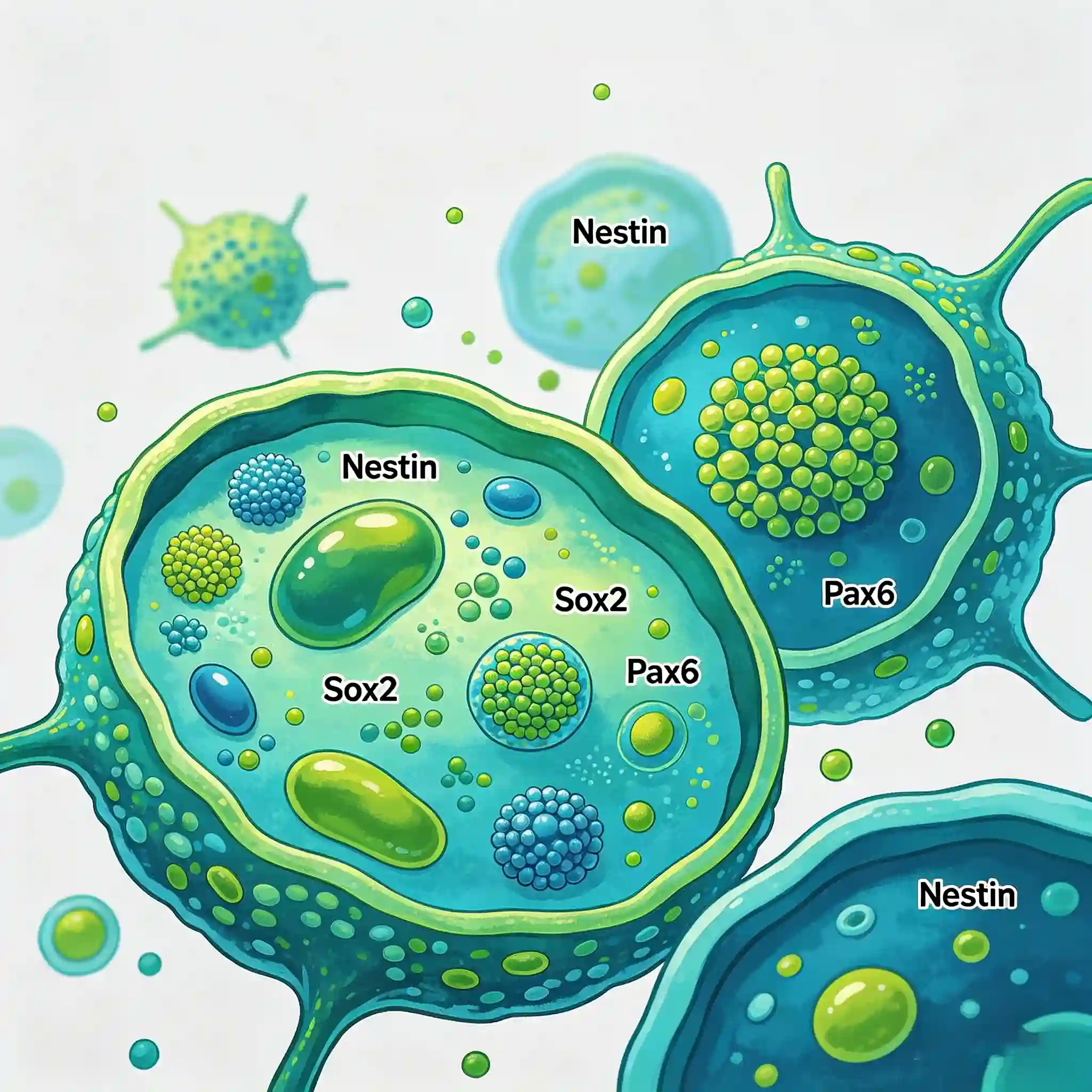
Neuroectodermal markers are a class of biomolecules that are specifically expressed in cells derived from the embryonic neuroectoderm, including proteins, enzymes, receptors, and transcription factors. They are primarily expressed in neurons, glial cells, neuroendocrine cells, neural crest cells, and their corresponding tumor cells.
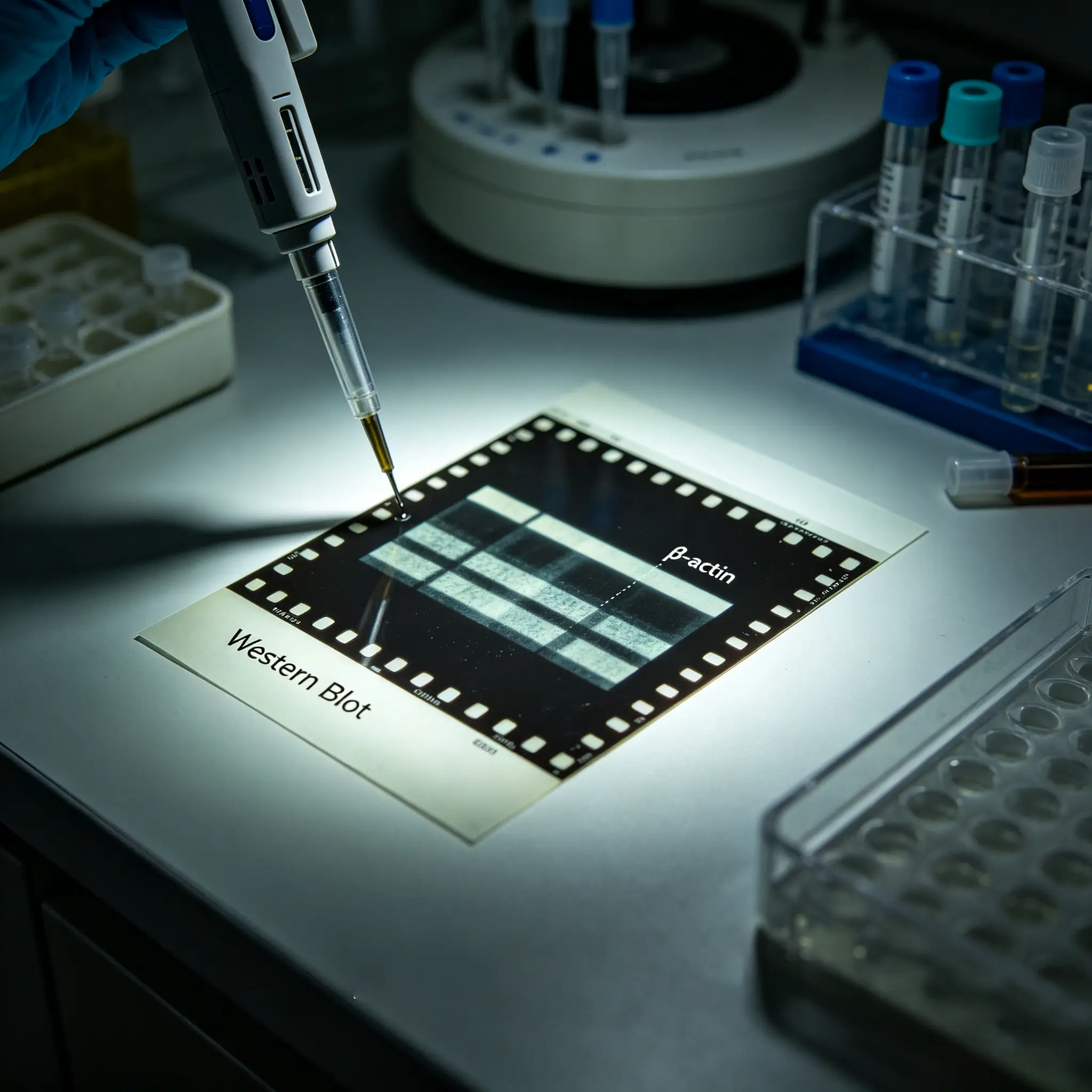
There is no "universal loading control", only the most suitable loading control for your experimental conditions. The key is to make a reasonable selection based on your cell line/tissue source, treatment conditions, and the molecular weight of the target protein.
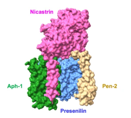
The γ secretase protease complex interacts with and cleaves intramembrane substrates as an essential function for regulation of intracellular signaling and cell-cell interactions. This multiprotein complex is comprised of four integral membrane proteins, Presenilin, Nicastrin, Aph-1, and PEN2, all of which are essential for complete proteolytic activity (1). Presenilin 1 and presenilin 2 are transmembrane proteins belonging to the presenilin family.
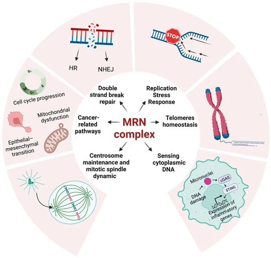
The Mre11-Rad50-Nbs1 (MRN) complex is a critical sensor and regulator in DNA double-strand break repair and genome stability maintenance. It participates in DNA damage repair pathways, checkpoint signaling, and ATM activation, and is closely related to embryonic development, immune cell maturation, and human genetic diseases. This paper introduces the molecular functions of the MRN complex and provides specific antibodies targeting its key components for related experimental research.
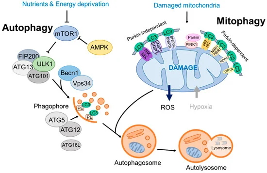
Mitophagy, a pivotal form of selective autophagy, is responsible for the specific elimination of damaged mitochondria to maintain mitochondrial homeostasis and cellular physiological functions. It is regulated by multiple conservative signaling pathways, including the FUNDC1 pathway, BNIP3/BNIP3L pathway, and PINK1/Parkin pathway, which are involved in various physiological and pathological processes such as hypoxia adaptation, erythroid cell maturation, and neurodegenerative diseases. This paper systematically elaborates on the core molecular mechanisms of mitophagy and collates a panel of high-quality antibodies targeting key proteins of mitophagy (e.g., LC3B, PINK1, Parkin, p62/SQSTM1, BNIP3). These antibodies are applicable to multiple experimental techniques including Western Blot, immunofluorescence, immunohistochemistry and immunoprecipitation, providing reliable molecular tools for the in-depth study of mitophagy-related biological processes and disease mechanisms.
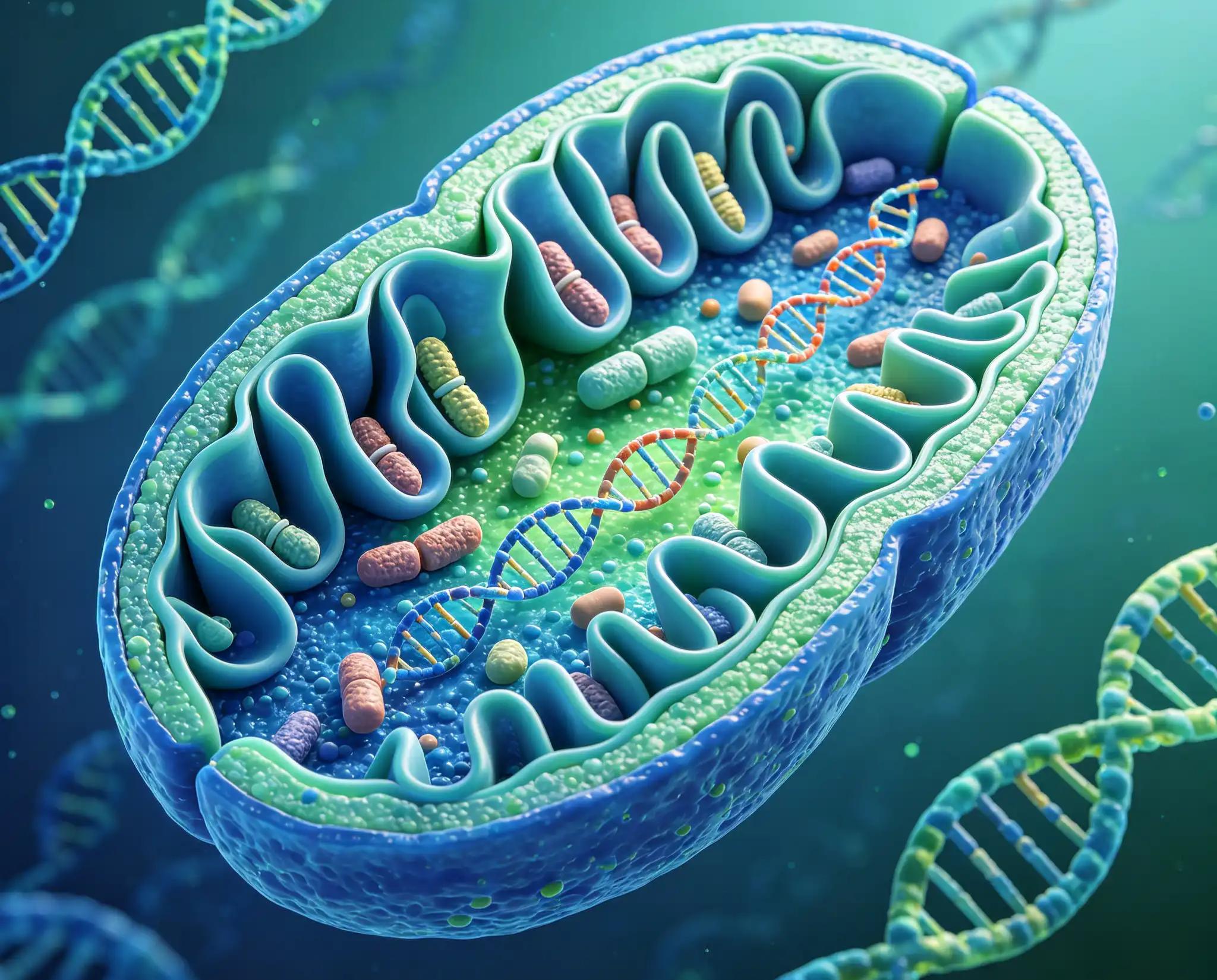
This study focuses on the Mitochondrial Marker Antibody Sampler Kit, which contains a variety of antibodies targeting classic mitochondrial proteins, including key mitochondrial proteins such as cytochrome c oxidase, cytochrome c, HSP60, prohibitin, pyruvate dehydrogenase complex, succinate dehydrogenase, SOD1, and voltage-dependent anion channel. These proteins are located in different substructures such as the inner mitochondrial membrane, intermembrane space, matrix, and outer membrane, and participate in core life activities including mitochondrial electron transport chain, tricarboxylic acid cycle, protein folding and stability, and metabolite transport. This paper systematically presents the product information of 8 rabbit monoclonal antibodies targeting the above mitochondrial proteins, as well as 2 matched secondary antibodies and internal reference antibodies, including catalog numbers, reactive species, and applicable experimental types.