


mTOR Pathway
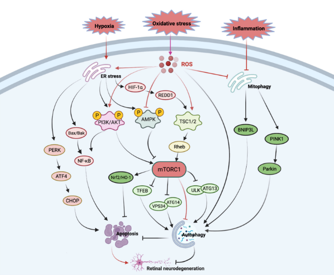
The mammalian target of rapamycin (mTOR) is a serine/threonine protein kinase that acts as a sensor of ATP and amino acids, coordinating nutrient availability and cell growth. Activated mTOR promotes the phosphorylation of p70 S6 kinase and inactivates 4E-BP1 through the PI3K/Akt signaling pathway, thereby regulating specific mRNA translation. mTOR forms two distinct complexes, mTORC1 (bound to Raptor) and mTORC2 (bound to Rictor and GβL), which mediate different downstream signaling events. Rapamycin inhibits mTORC1 activity by disrupting the mTOR–Raptor interaction. Abnormal regulation of mTOR is closely associated with tumorigenesis and ocular neurodegenerative diseases, making it a promising therapeutic target. This paper reviews the core molecular mechanisms of the mTOR signaling pathway and its relevance to retinal neurodegeneration, providing a theoretical basis for targeted antibody development and disease intervention.
Mediator Complex
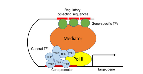
The mammalian Mediator complex is a multi-subunit protein complex that relays signals from transcription factors to RNA polymerase II (Pol II) and the basal transcription machinery, acting as a centralized hub and integrator for transcriptional regulation. It participates in epigenetic regulation, transcriptional elongation, transcription termination, mRNA processing, non-coding RNA activation, and super-enhancer formation, thereby regulating insulin signaling, NF-κB-dependent signaling, stem cell pluripotency and self-renewal, colon cancer cell proliferation and other key biological processes. The mammalian Mediator complex consists of 31 protein subunits and can be divided into four structural modules: head, middle, tail, and CDK8 kinase module. The CDK8 kinase module acts as a molecular switch, phosphorylating the C-terminal domain of Rpb1, the large subunit of RNA polymerase II, to inhibit the recruitment and transcription initiation of RNA polymerase II.
Mature Neuron Marker
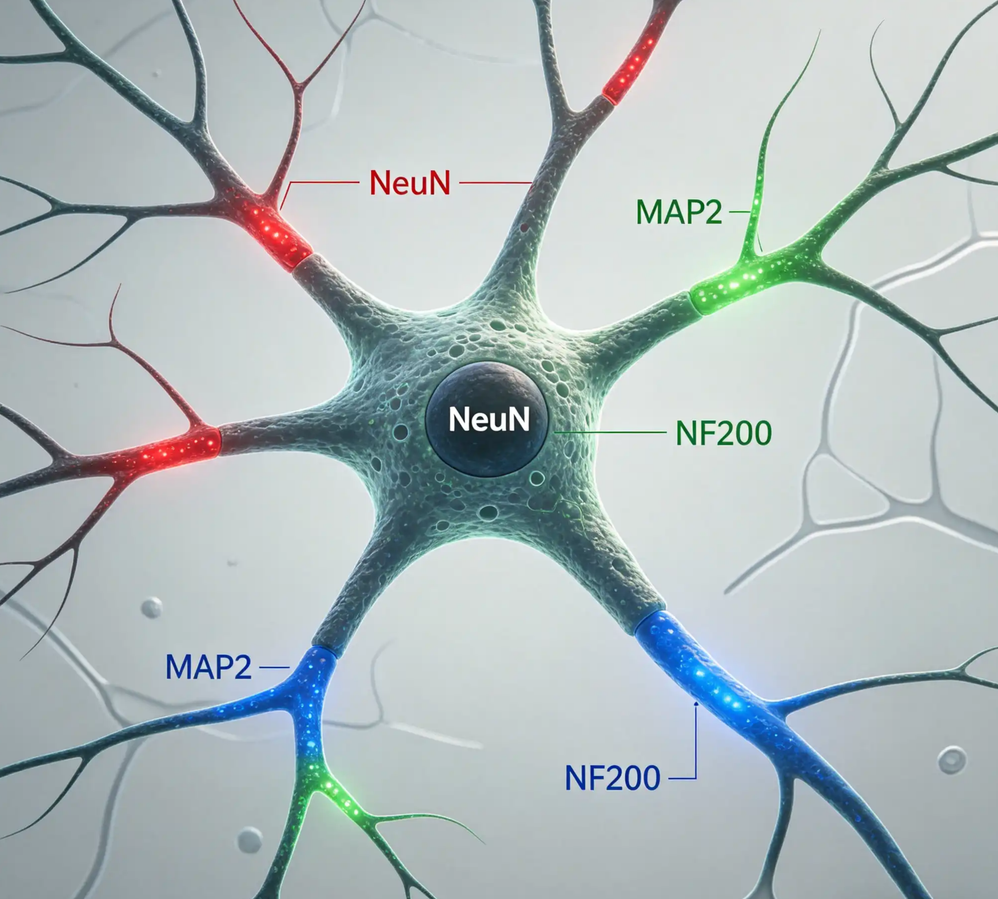
This study focuses on the identification and characterization of mature neuron markers, and systematically elaborates the expression characteristics and biological functions of a variety of specific markers in mature neurons. As a core nuclear protein, NeuN is widely expressed in most post-mitotic neurons; GAP43, β3-tubulin, MAP2 and neurofilament protein (NEFL) participate in neuronal process growth, cytoskeleton construction and axon development; Synaptophysin and PSD95 regulate the structural and functional functions of presynaptic and postsynaptic respectively, and UCH-L1 maintains normal synaptic function of neurons through deubiquitination. At the same time, the information of specific antibodies that can be used to detect the above markers is integrated, and their reactive species and application scenarios are clarified, providing theoretical basis and experimental reference for the identification of mature neurons, the study of neurobiological mechanisms and the application of related antibody reagents.
Matrix Remodeling
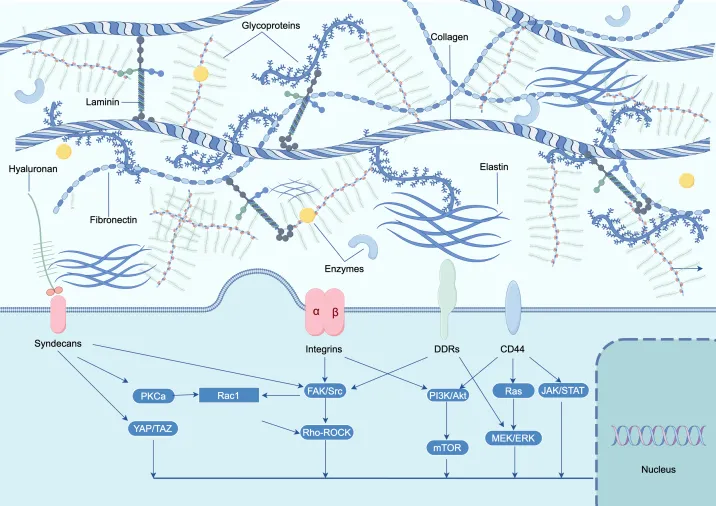
Matrix remodeling is a critical physiological process that maintains tissue homeostasis and regulates cellular biological functions, which is precisely controlled by matrix metalloproteinases (MMPs) and their endogenous tissue inhibitors of metalloproteinases (TIMPs). As a family of zinc-dependent proteases, MMPs target and degrade various extracellular matrix proteins, and participate in embryonic development, wound healing, tumor invasion, angiogenesis, carcinogenesis, and apoptosis. The activity of MMPs is regulated by transcriptional control and post-translational cleavage. TIMPs can irreversibly inhibit the protease activity of MMPs by direct binding and chelating the zinc cofactor at the catalytic site. This study summarizes the antibody products targeting key molecules including MMP-2, MMP-3, MMP-9, TIMP1-3, clarifies their reactivity and application scenarios, and provides reliable antibody tools and experimental references for basic research related to matrix remodeling.
YAP/TAZ Transcriptional Targets Antibody
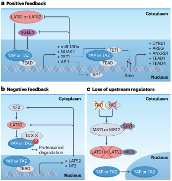
YAP and TAZ (WWTR1) are transcriptional co-activators that play a central role in the Hippo Signaling pathway that regulates cell, tissue and organ growth. Under growth conditions, YAP and TAZ are translocated to the nucleus, where they interact with DNA-binding transcription factors (e.g., Transcriptional Enhanced Activation Domain [TEAD] proteins) to regulate the expression of genes that control fundamental aspects of cell function, such as proliferation and cell survival (1). A number of genes have been experimentally confirmed as targets of transcriptional regulation by YAP and TAZ. These include the extracellular matrix proteins CTGF, CYR61, and integrin β2 (2-4), the inhibitor of apoptosis protein (IAP) survivin (5), the mechano-sensitive nuclear envelope protein Lamin B2 (6), and the oncogenic receptor tyrosine kinase Axl (7).
Literature Sharing: Microglia-specific Regulation of Lipid Metabolism in Alzheimer's Disease
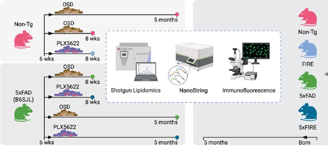
Literature Sharing: Microglia-specific Regulation of Lipid Metabolism in Alzheimer's Disease I. Research Background Alzheimer's disease (AD), as the most common neurodegenerative disease and major cause of dementia, is characterized by severe disruption of brain homeostasis as one of its core pathological features. The role of abnormal lipid metabolism in disease progression has long been insufficiently recognized. In recent years, with the development of lipidomics technology, studies have found that imbalances in lipids such as phospholipids, sphingolipids, and cholesterol may be initiating factors in AD pathogenesis, rather than mere pathological byproducts. These lipid disorders can promote amyloidogenesis through mechanisms such as regulating membrane fluidity, secretase compartmentalization, and Aβ aggregation kinetics. The study "Microglia-specific regulation of lipid metabolism in Alzheimer's disease revealed by microglial depletion in 5xFAD Mice" focuses on microglia—the brain's resident immune cells. Combining the 5xFAD transgenic mouse model with human post-mortem brain tissue samples, the study investigates the specific regulatory role of microglia in lipid metabolism disorders in AD. The aim is to decipher the association between abnormal lipid metabolism in AD and different brain cell types and molecular pathways, providing new therapeutic targets for the disease. Previous studies have confirmed that lipid metabolism risk genes enriched in microglia (such as TREM2, PLCG2, GRN, etc.) are involved in Aβ clearance and neuroinflammation regulation. However, the specific impact of microglial depletion on brain lipid metabolism has been widely overlooked, with only one study focusing on its necessity for leukotriene synthesis, which became the core entry point for this study.
β Catenin Antibody
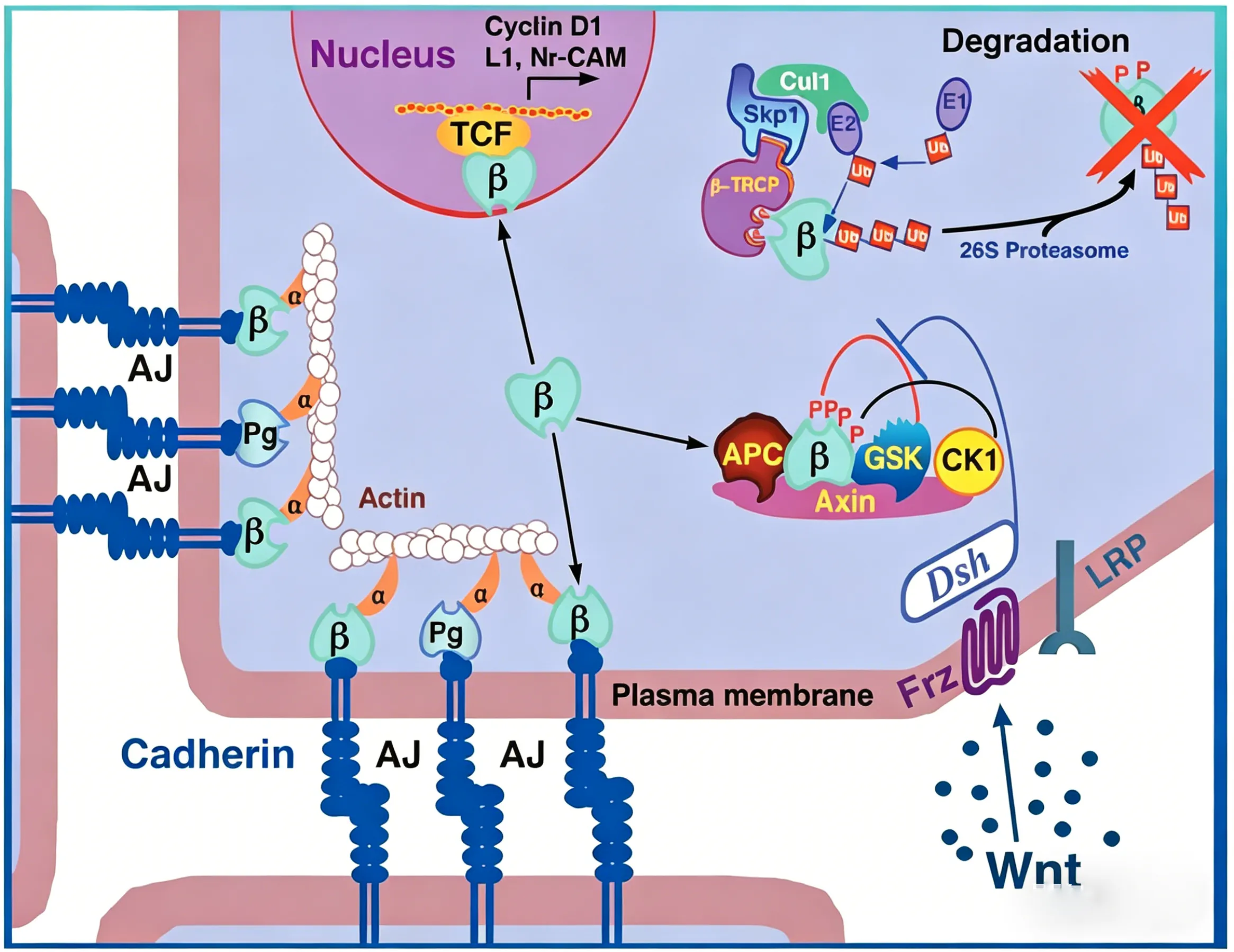
β-catenin is a key downstream effector in the Wnt signaling pathway (1). It is implicated in two major biological processes in vertebrates: early embryonic development (2) and tumorigenesis (3). CK1 phosphorylates β-catenin at Ser45. This phosphorylation event primes β-catenin for subsequent phosphorylation by GSK-3β (4-6). GSK-3β destabilizes β-catenin by phosphorylating it at Ser33, Ser37, and Thr41 (7). Mutations at these sites result in the stabilization of β-catenin protein levels and have been found in many tumor cell lines (8).