
Literature Sharing: Multiplex Immunofluorescence – A New Approach for Tumor Immune Cell Atlas Detection
I. Research Background
In the era of rapid development of tumor immunotherapy, precise analysis of the tumor immune microenvironment and identification of specific immune biomarkers are crucial for improving the prognostic prediction of immunotherapy and optimizing personalized treatment strategies. In 2021, Hernandez et al. published a study entitled "Multiplex Immunofluorescence Tyramide Signal Amplification for Immune Cell Profiling of Paraffin-Embedded Tumor Tissues" in Frontiers in Molecular Biosciences, focusing on the technical challenges of precise immune cell classification and atlas construction in tumor tissues. Previous clinical and basic research has confirmed that the density, phenotype, and spatial distribution of immune cells in the tumor immune microenvironment directly determine tumor development, invasion, metastasis, and patient response to immunotherapy, serving as the core basis for evaluating tumor prognosis and screening therapeutic targets. Traditional immunohistochemistry (IHC) technology can only detect a single marker in a single sample, which not only consumes a large amount of clinically scarce paraffin-embedded tumor tissue samples and has limited detection parameters but also tends to have problems such as signal overlap and insufficient detection accuracy. It cannot achieve simultaneous analysis of multiple immune cell phenotypes and multiple checkpoint proteins, nor can it restore the spatial interaction relationships between cells in the tumor microenvironment, which greatly limits the research on tumor immune mechanisms and the development of new immunotherapies. Based on this, this study introduces tyramide signal amplification (TSA) multiplex immunofluorescence (mIF) technology, aiming to break through the limitations of traditional detection techniques and establish a standardized technical system that can achieve simultaneous detection of multiple markers, precise immune cell classification, and spatial atlas analysis on a single paraffin-embedded tumor tissue section, providing reliable technical support for in-depth research on the tumor immune microenvironment, immune biomarker discovery, and the implementation of precision immunotherapy.
II. Research Methods
This study used formaldehyde-fixed, paraffin-embedded multi-type tumor tissues as research objects to construct a multiplex immunofluorescence detection system adapted to different tumor subtypes and research targets. The overall process covers experimental system design, sample screening, image segmentation analysis, marker interpretation, and data integration and validation. Combining multidisciplinary expertise in oncology, pathology, and immunology, the research team customized differentiated mIF detection panels based on research hypotheses, target cell populations, and therapeutic research directions. Up to 8 markers can be simultaneously labeled on a single tissue section, while specific epithelial, stromal, and tumor markers are matched for different tumor types to accurately distinguish cells from different tumor tissue origins. In terms of sample screening, strict criteria were set for tissue sample size, tumor cell proportion, and effective malignant cell count. Samples with necrotic areas, mucus interference, and other disqualifications were excluded, and the reliability of the experimental foundation was ensured through pathological quality assessment. The experiment relied on Inform image analysis software to complete tumor region division, tissue and single-cell segmentation. Combined with DAPI nuclear staining technology, parameters such as nuclear size and staining threshold were optimized to achieve precise single-cell localization. The study distinguished specific cell phenotypes by identifying subcellular localization expression characteristics of different markers, and excluded tissue autofluorescence interference with negative controls. Finally, integrating data analysis tools such as R-studio and SAS, combined with cell XY coordinate information, co-expression analysis of markers, immune cell classification, cell density statistics, and spatial distribution feature analysis were completed. Meanwhile, through multi-panel shared internal reference markers and matched section cutting levels, the comparability and accuracy of multiple groups of experimental data were ensured.
III. Results Analysis
3.1 Precision Classification Effect of Tumor-Specific Markers
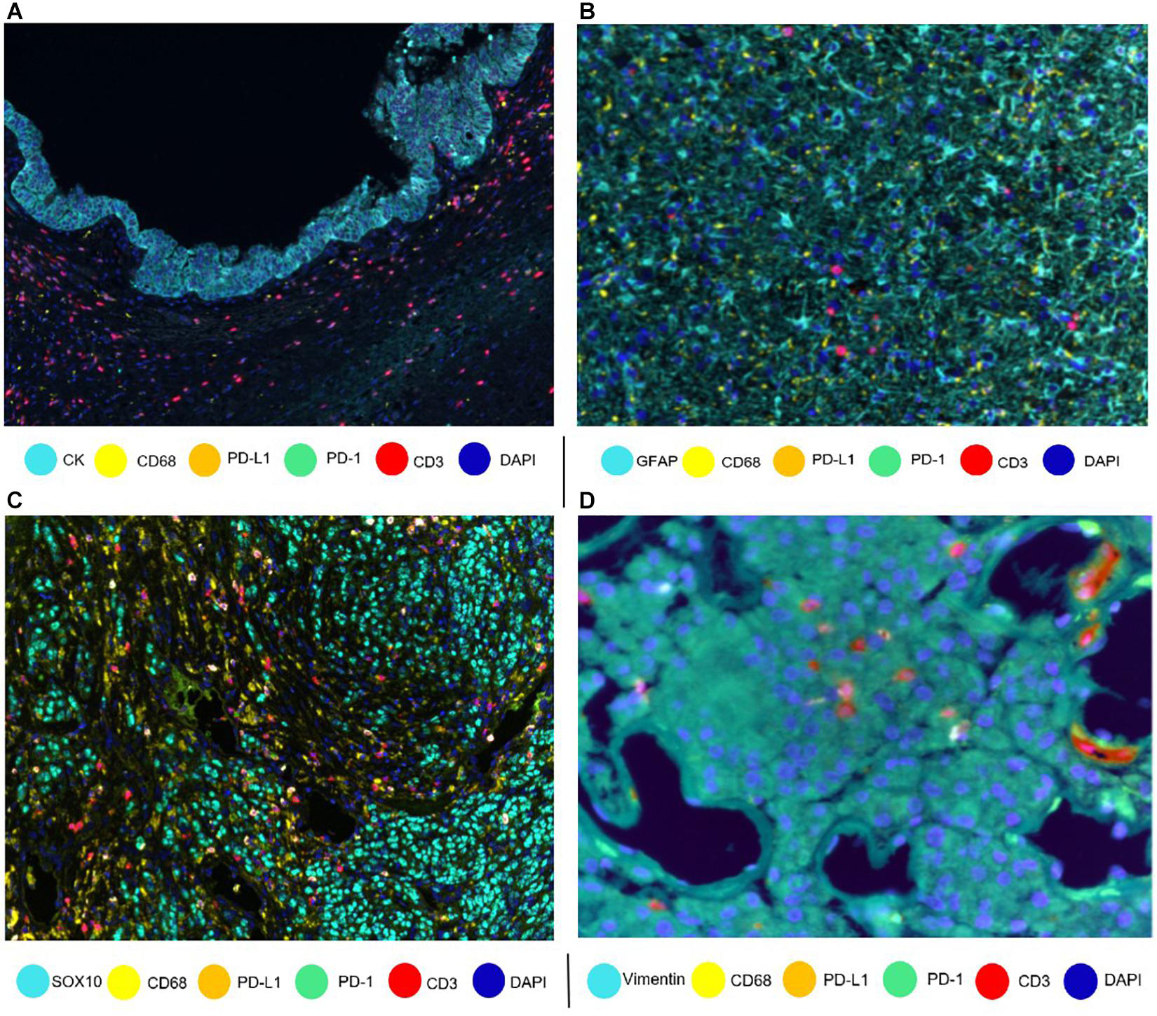
Based on the customized multiplex immunofluorescence marker detection system, this study conducted specific cell classification verification for multiple types of solid tumors, effectively solving the industry pain point that different tumor cells have high morphological similarity and are difficult to accurately identify by conventional staining techniques. The study matched exclusive specific markers for four typical tumor tissues. In pancreatic intraductal papillary mucinous tumor tissues, cytokeratin (CK) accurately labeled epithelial-derived tumor cells, clearly outlining the boundary of tumor epithelial tissue structure; in glioblastoma tissues, glial fibrillary acidic protein (GFAP) specifically targeted glial-derived tumor cells, achieving clear segmentation of tumor cells from surrounding normal brain tissue and infiltrating immune cells; melanoma tissues relied on SOX10 nuclear-specific expression characteristics to accurately locate melanoma tumor cells, and the nuclear-localized marker expression pattern further improved the classification accuracy; while sarcoma tissues clearly distinguished sarcoma stromal-derived tumor cells through specific labeling of vimentin. From the imaging results, all types of markers showed no obvious non-specific staining, with clear target cell labeling signals and distinct boundaries, fully verifying that mIF technology can flexibly customize detection panels according to tumor tissue origin and pathological type, and can adapt to the cell classification needs of different solid tumors, laying a precise cell localization foundation for subsequent differential analysis of immune atlases in different tumor microenvironments.
3.2 Construction of Immune Cell Phenotype Algorithm and Precision Classification in Tumor Tissues
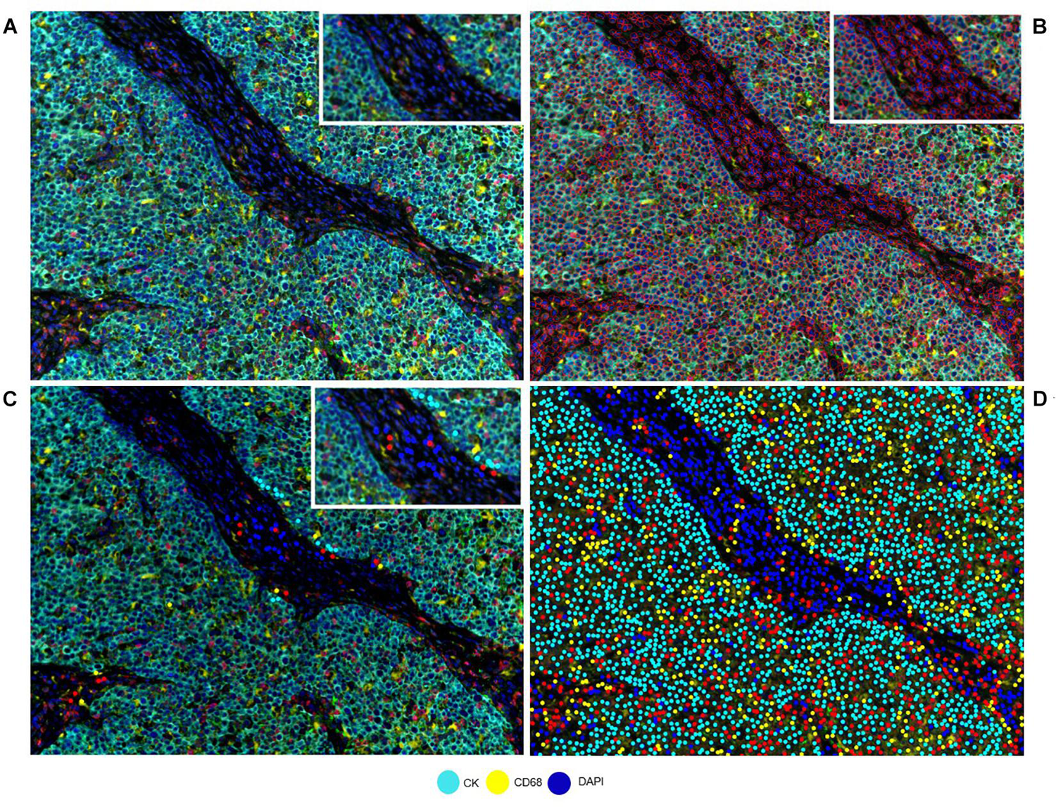
To solve the problem of standardization and precision in tumor tissue immune classification, the study combined multiplex immunofluorescence technology with intelligent image analysis algorithms to build a complete analysis system from tissue imaging to cell phenotype identification, fully demonstrating the application advantages of this technology in complex tumor pathological samples. Taking pancreatic intraductal papillary mucinous tumor tissue as a model, the study first obtained a complete tissue composite image through multiple marker overlay imaging, comprehensively covering the marker expression information of tumor cells, stromal cells, and infiltrating immune cells; then relied on image analysis software to complete automatic cell segmentation, accurately outlining the contour of each single cell with red lines, achieving precise disassembly from tissue level to single-cell level, avoiding classification errors caused by cell adhesion and blurred boundaries in traditional detection. During the algorithm training process, the study continuously optimized classification parameters by combining pathological morphological features and marker expression rules, forming standardized cell phenotype determination rules. The final output classification results can accurately distinguish the marker expression profiles of different cells, clearly identifying the phenotypic differences between various immune cells and tumor cells. The entire process achieved a standardized closed loop from original imaging, cell segmentation to intelligent classification, solving the problems of strong subjectivity in manual interpretation and single classification in traditional technology, providing mature algorithm support for automated and precise analysis of immune phenotypes in large batches of tumor tissues.
3.3 Co-expression Characteristics of Immune Cells in Non-Small Cell Lung Cancer Tumor Microenvironment
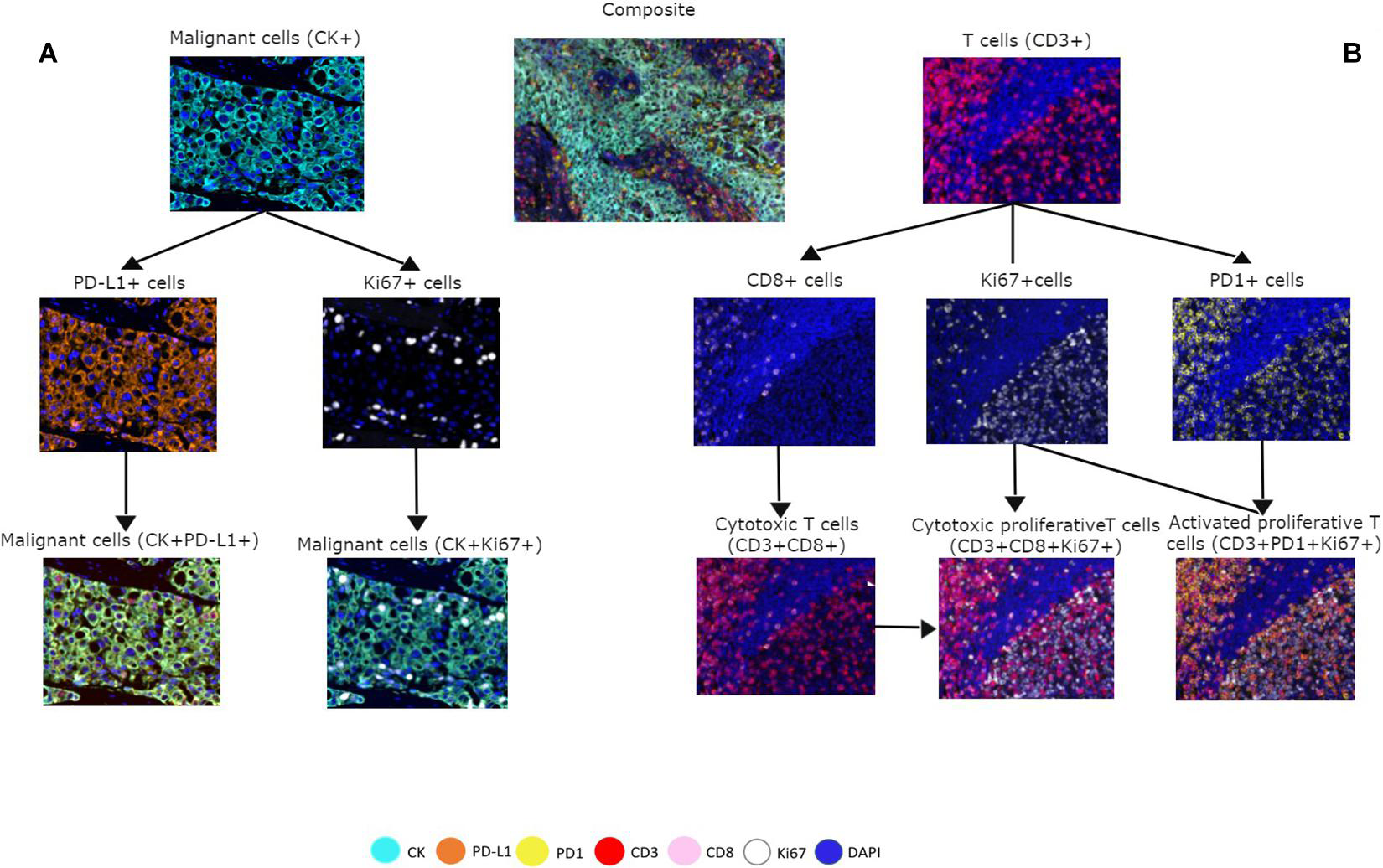
With the core advantage of mIF technology in simultaneous detection of multiple targets, the study deeply analyzed the cell interaction patterns in the non-small cell lung cancer tumor microenvironment, clearly restoring the marker co-expression characteristics and spatial distribution patterns of tumor cells and multiple immune cells, intuitively revealing the complex regulatory mechanisms of the lung cancer immune microenvironment. At the tumor cell level, CK-labeled malignant tumor cells can simultaneously detect the co-expression of PD-L1 and Ki67 markers, intuitively confirming that tumor cells have immune escape potential and proliferative activity, explaining the core mechanism of continuous lung cancer progression and resistance to immune killing. At the immune cell level, the detection system can accurately distinguish the functional phenotypes of multiple T cell subsets: CD3+CD8+ co-expressing cells are classic cytotoxic T cells, which are the core effector cells of the body's anti-tumor immunity; Ki67+CD3+CD8+ co-expressing proliferative cytotoxic T cells reflect active anti-tumor immune responses in the tumor local area; while CD3+Ki67+PD-1+ co-expressing activated exhausted T cells accurately capture the key characteristics of immune cell functional exhaustion and immune suppression formation in the tumor microenvironment. These multi-dimensional and refined cell phenotype information cannot be simultaneously obtained by traditional single-label IHC technology. It not only clearly shows the complex state of coexistence of immune activation and immune suppression in the lung cancer tumor microenvironment but also provides intuitive cytological evidence for analyzing lung cancer immune escape mechanisms and screening effective immunotherapy targets.
3.4 Distribution Characteristics of Specific Immune Cell Subsets in Liver Cancer Tissues
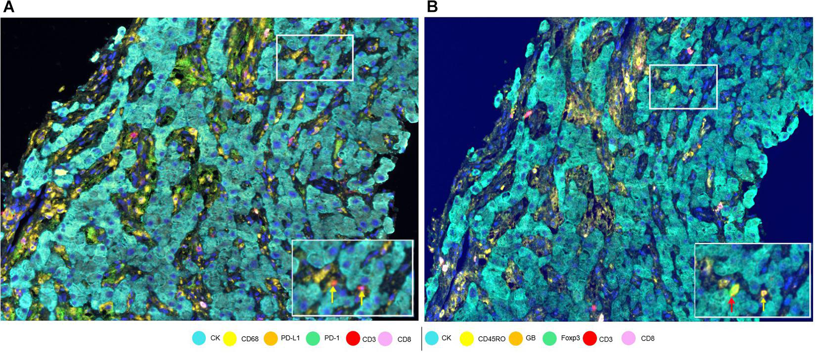
Aiming at the research difficulty of sparse immune infiltration and difficulty in identifying rare immune cells in the liver cancer microenvironment, this study utilized the ultra-high signal amplification capability of mIF-TSA to achieve precise capture of weak immune signals and refined classification of rare T cell subsets, compensating for the inherent defects of traditional detection techniques. Under low-power magnification, a small number of infiltrating CD3+ T cells can be clearly located in the tissue, confirming the pathological feature of low immune infiltration level in the liver cancer tumor microenvironment; after high-power magnification imaging, two T cell subsets with distinct functions can be accurately distinguished: one is CD3+CD45Ro+ memory T cells, which can persist in the tumor microenvironment for a long time and participate in the construction of local immune memory; the other is CD3+Foxp3+CD45Ro+ regulatory T cells, as core immune-suppressive cells, they can block the anti-tumor effect of effector T cells by secreting inhibitory cytokines, which is a key inducement for the formation of the immune-suppressive microenvironment in liver cancer. Traditional detection techniques are difficult to distinguish such rare T cell subsets with similar morphology but significant functional differences, while mIF technology achieves precise classification and spatial localization of rare immune cells through multi-marker co-localization analysis, clearly revealing the functional heterogeneity of a small number of infiltrating immune cells in the liver cancer microenvironment, providing important theoretical basis for explaining the clinical phenomena of low immunotherapy response rate and large prognostic differences in liver cancer.
3.5 OX40 Activation Characteristics in Non-Small Cell Lung Cancer Tumor Cells
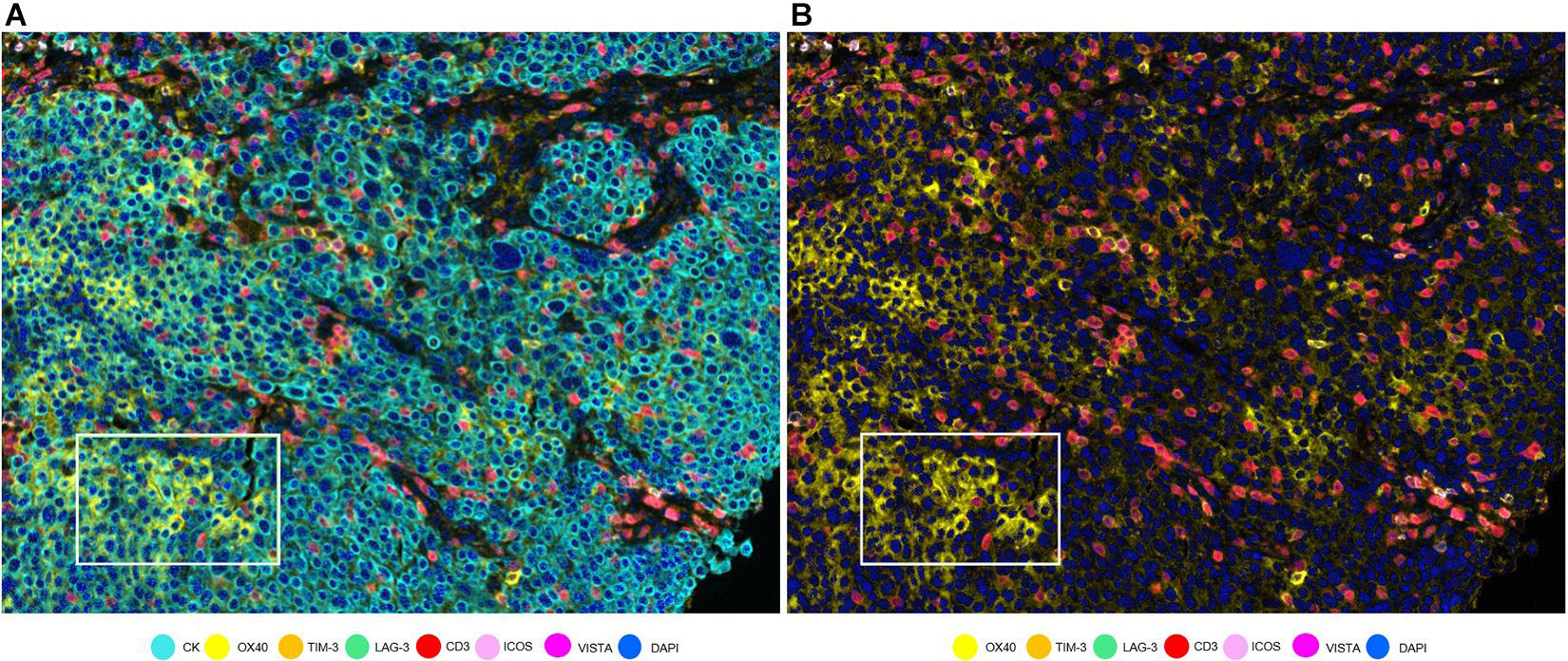
Relying on the technical characteristics of multi-marker co-localization analysis, this study broke through the limitations of traditional immune research, no longer focusing solely on infiltrating immune cells, and successfully discovered the expression characteristics of a new immune activation target in non-small cell lung cancer tumor cells, providing a new idea for tumor immune target discovery. Through multi-marker co-localization analysis, specific co-expression of CK and OX40 was detected in non-small cell lung cancer tumor cells. As a key immune co-stimulatory molecule, the expression of OX40 on the surface of tumor cells suggests that tumor cells have the potential to actively regulate local immune responses. In the imaging results, single imaging after removing the CK marker can clearly highlight the OX40-positive expression area, excluding the signal interference of immune cell markers, and accurately verifying the expression characteristics of tumor cell-derived OX40. This discovery breaks the traditional cognition that OX40 is only expressed in immune cells, confirming that tumor cells can participate in the regulation of the tumor immune microenvironment by expressing co-stimulatory molecules, providing new experimental evidence for the development of new immune targets for lung cancer and the design of combined immunotherapy regimens, fully reflecting the unique value of mIF technology in discovering new tumor immune markers.
3.6 Comparison of Detection Effects of Glioblastoma by Multiple Technical Systems
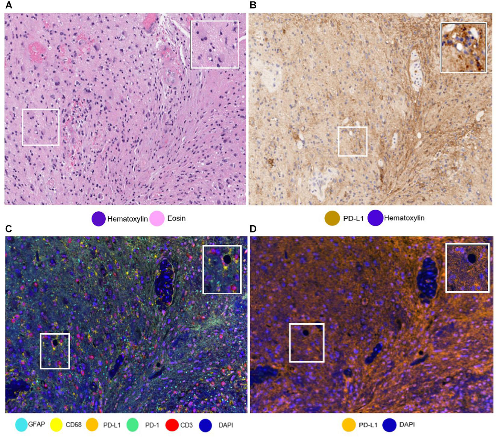
To intuitively verify the application value of mIF technology in the detection of difficult tumors, the study simultaneously used three detection methods: HE staining, traditional IHC, and multiplex immunofluorescence to detect glioblastoma samples, clearly highlighting the unique advantages and clinical practicality of multiplex fluorescence technology. Conventional HE staining can only clearly show the pathological morphological structure of tumor tissue, cannot distinguish infiltrating lymphocytes from glioblastoma tumor cells, and is difficult to carry out immune microenvironment analysis; traditional single-label PD-L1 IHC staining can only show PD-L1 positive signals alone, but cannot distinguish whether the signals come from tumor cells, macrophages, or lymphocytes, which is very easy to lead to misinterpretation of results. The complex characteristics of PD-L1-expressing cell types in glioblastoma further amplify the detection defects of traditional technology. In contrast, the mIF multi-staining system can accurately distinguish between the two types of cells by using GFAP to label glioblastoma cells and CD3 to label infiltrating lymphocytes. At the same time, separate PD-L1 signal imaging can accurately locate the cellular source of PD-L1, completely avoiding the interpretation errors of traditional technology. This comparison fully proves that mIF technology can accurately solve the immune marker detection problems of difficult tumor types through multi-marker co-localization classification, providing a standardized detection scheme for immune marker evaluation and immunotherapy efficacy prediction of refractory tumors such as glioblastoma.
IV. Conclusion
This study systematically constructed a multiplex immunofluorescence detection technology system based on tyramide signal amplification, successfully breaking through many bottlenecks of traditional single immunohistochemistry technology in tumor immune microenvironment research, providing a mature and reliable technical paradigm for tumor immune cell atlas mapping, immune biomarker discovery, and immunotherapy mechanism exploration. Compared with the defects of traditional IHC technology that can only detect single markers, has large sample consumption, and cannot analyze cell spatial interactions, the optimized mIF-TSA technology in this study can complete simultaneous detection of up to 8 markers on a single paraffin-embedded tumor section, with ultra-high detection sensitivity and classification accuracy. It can not only accurately distinguish tumor cells of different pathological types such as lung cancer, liver cancer, glioma, and sarcoma but also refine the classification of various functionally heterogeneous immune cell subsets, effectively capturing rare immune cells, weak marker signals, and cell co-expression characteristics that are difficult to identify by traditional technology. At the same time, combined with digital image analysis and spatial statistical methods, this technology breaks through the limitation of single cell density statistics, can accurately analyze the spatial distribution and interaction relationships between tumor cells and immune cells, clearly reveal the differentiated patterns of immune activation and immune suppression in different tumor microenvironments, and can also discover new tumor immune targets such as OX40, greatly expanding the depth and breadth of tumor immune research. However, this technology still has certain limitations. The tyramide signal amplification process is prone to signal overload umbrella effect, affecting detection accuracy. The entire experiment and data analysis process takes longer and requires high pathological professional literacy and data analysis capabilities of operators. Moreover, the number of fluorescence spectra limits the number of markers detected in a single panel to a certain extent. Overall, with its core advantages of multi-dimensionality, high precision, and preservation of tissue spatial information, mIF-TSA technology effectively compensates for the shortcomings of traditional tumor immune detection technology, providing strong technical support for tumor prognosis assessment, immunotherapy efficacy prediction, and new immunotherapy development, and has extremely high application value and broad development prospects in the field of tumor precision immunology research and clinical translation.
References
Hernandez S, Rojas F, Laberiano C, Lazcano R, Wistuba I and Parra ER (2021) Multiplex Immunofluorescence Tyramide Signal Amplification for Immune Cell Profiling of Paraffin-Embedded Tumor Tissues. Front. Mol. Biosci. 8:667067. doi: 10.3389/fmolb.2021.667067.
EnkiLife not only provides customers with a complete set of TSA multiplex labeling kits, but also offers various TSA specialty technical services, including IF fluorescence staining, fluorescence panoramic scanning, ultra-multiplex staining, and pathological analysis (5 markers and below).
Product | Catalog Number |
|---|---|
TSA Six-Label Seven-Color Multiplex Immunohistochemistry Kit | |
TSA Five-Label Six-Color Multiplex Immunohistochemistry Kit | |
TSA Four-Label Five-Color Multiplex Immunohistochemistry Kit | |
TSA Three-Label Four-Color Multiplex Immunohistochemistry Kit | |
TSA Two-Label Three-Color Multiplex Immunohistochemistry Kit |
For details, please check TSA mIHC Kit