
Visualizing Why TSA Technology Outperforms Traditional IHC/IF Through Experimental Images
Tyramide Signal Amplification Technology: Hardcore Advantages Compared with Literature Experimental Data, Professional Analysis of Biological Research Tissue Staining
In immunohistochemistry (IHC) and immunofluorescence (IF) experiments, many researchers face the same challenges: weak signals, high background, failed multi-labeling, and poor result reproducibility. Traditional IHC relies on chromogenic substrates for qualitative detection, while conventional IF depends on direct fluorescent labeling. Although seemingly mature, these methods have insurmountable technical bottlenecks in detecting low-abundance targets, co-localizing multiple markers, and adapting to diverse sample types.
Most technical optimizations on the market are merely cosmetic fixes that fail to address the root cause. In contrast, TSA (Tyramide Signal Amplification) technology achieves comprehensive breakthroughs through its unique iterative staining and signal amplification system. The emergence of TSA has completely broken the limitations of traditional staining techniques. This article precisely dissects the exclusive advantages of TSA over traditional IHC/IF from six core dimensions, combining experimental results from literature to comprehensively compare the differences between TSA and traditional IHC/IF, helping researchers accurately select the optimal experimental protocol and bid farewell to the frustration of repeated optimization attempts and failed results.
1. Sensitivity Overwhelms: The Only Staining System Capable of Stably Detecting Low-Abundance Targets
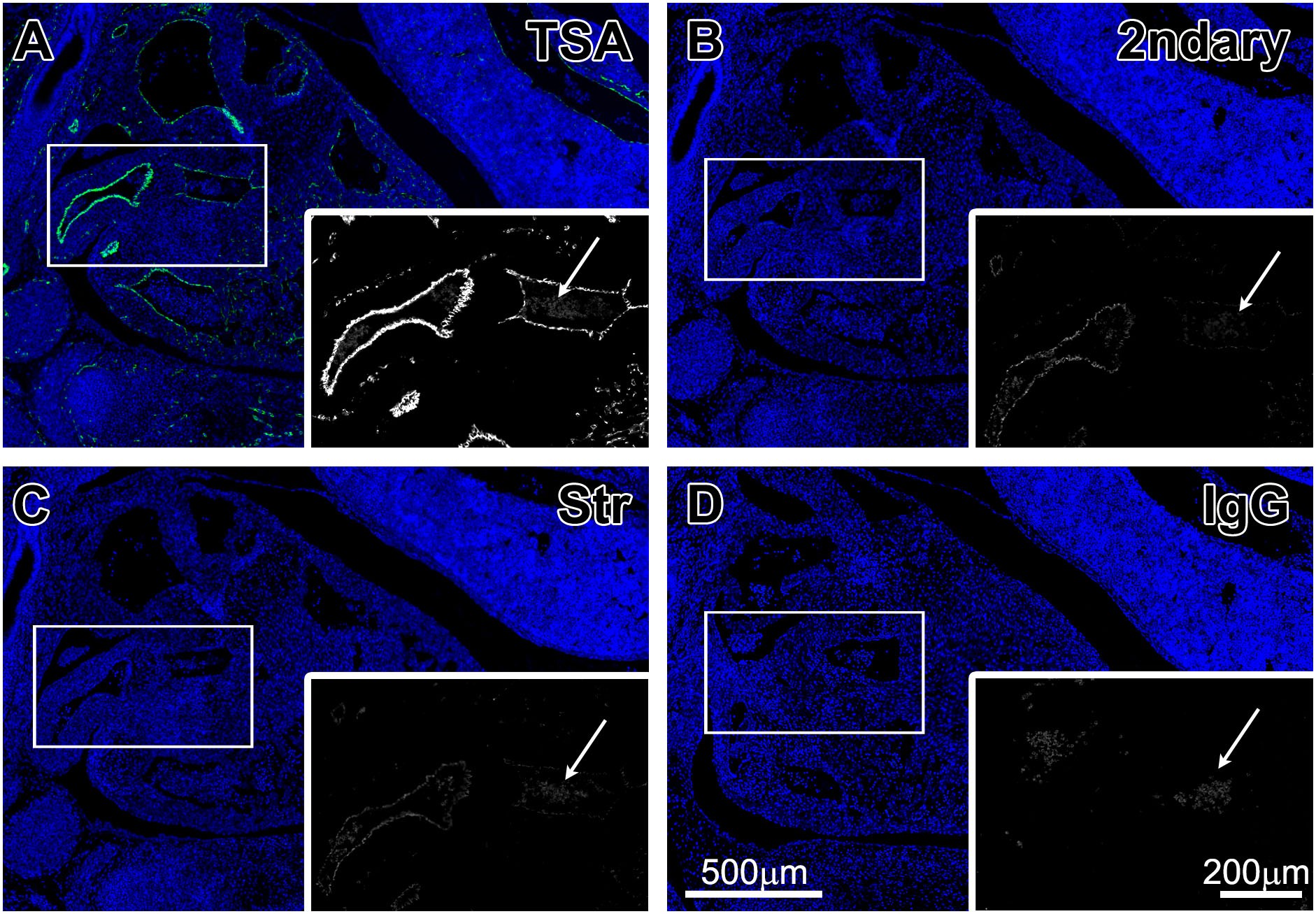
Multiple control experiments have intuitively confirmed the core shortcoming of traditional methods: two conventional approaches—direct fluorophore-conjugated secondary antibodies and streptavidin-direct fluorescence labeling—can only detect signals from highly expressed targets. For low-abundance, weakly expressed target proteins such as tissue microvessels, traditional methods completely lose signal, easily causing false-negative results that seriously affect the authenticity of experimental data.
In contrast, the TSA signal amplification system demonstrates absolute advantages: relying on HRP-catalyzed tyramide fluorogenic substrate to specifically deposit at antigen sites, achieving precise cascade signal amplification. In control experiments using the same mouse embryonic tissue and CD31 target, only TSA technology can clearly capture the weak specific signals of low-expressing microvessels. The signal hierarchy in high-expression regions is also clearer, with no additional non-specific background. This confirms that TSA is a reliable technology for stably detecting low-abundance targets, perfectly solving the defect of traditional technologies where "strong signals are visible, but weak signals are completely lost."
2. Precise Differentiation of Immune Cell Subsets: Clear Cell Subset Typing and Co-localization Validation
Traditional IHC/IF multi-labeling has significant drawbacks in immune cell typing research: multi-target staining tends to produce signal overlap and blurred boundaries, making it impossible to accurately distinguish different T cell subsets or clearly verify marker co-expression relationships, thus failing to meet the refined research needs of immune microenvironment and cell typing.
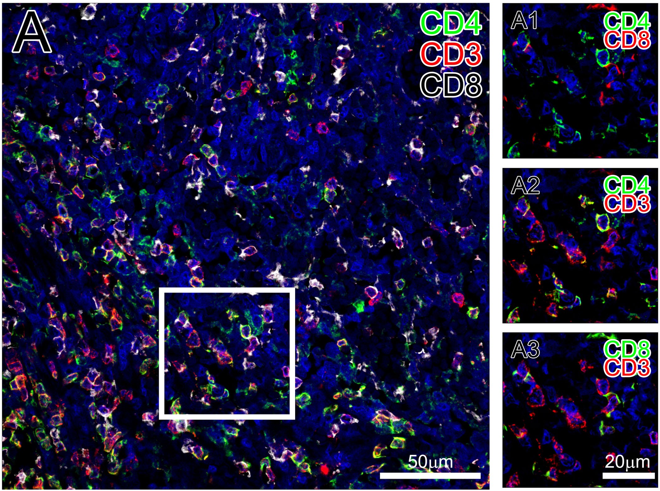
Multi-color staining results in human spleen tissue directly validate the multi-labeling advantages of TSA: through the TSA multiplex fluorescence system, triple-marker simultaneous staining of CD3, CD4, and CD8 was successfully achieved. The results clearly distinguish two mutually exclusive T cell subsets (CD4+ and CD8+), while precisely verifying that both subsets co-express the universal T cell marker CD3. Cell boundaries are clear, signals show no crosstalk, and the negative background is clean. Compared with the blurry multi-labeling effect of traditional technologies, TSA enables precise immune cell typing, target co-localization, and cell distribution analysis, adapting to various immune mechanism studies.
3. Anti-Nonspecific Interference: Effectively Avoiding Plasma Cell False Staining for More Accurate Results
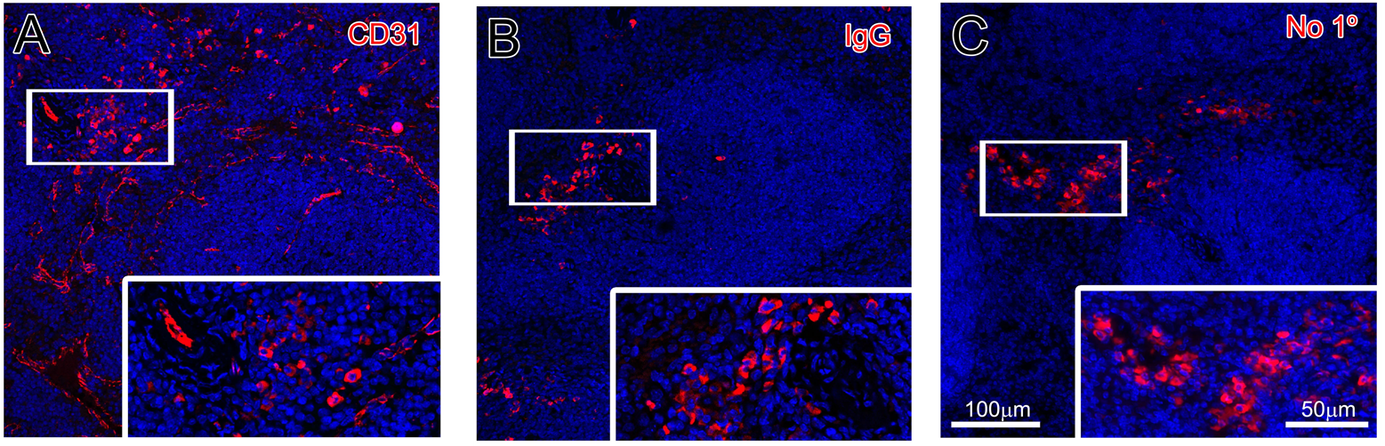
This is a critical pain point easily overlooked in traditional multi-label staining: mouse immune tissues (spleen) are rich in plasma cells, which highly express immunoglobulins. When using traditional staining methods, secondary antibodies will non-specifically bind to endogenous Ig in plasma cells, producing a large number of false-positive signals. Even with isotype controls or blank controls, non-specific staining persists, seriously interfering with the interpretation of real target signals and easily leading to misjudgment of experimental conclusions.
In contrast, the optimized TSA staining system, through precise site blocking and iterative staining procedures, minimizes the non-specific background caused by plasma cells. In the same mouse spleen tissue staining experiment, the TSA system can effectively distinguish real target signals from plasma cell false signals, significantly reducing the non-specific staining problem caused by endogenous immunoglobulins. This makes in-situ tissue staining results purer and interpretation more accurate—a key optimization unattainable by traditional staining systems.
4. Ultra-High Throughput Four-Color Labeling: Multi-Dimensional Analysis of Tumor Tissue Characteristics on a Single Section
Traditional IHC can only achieve dual-color labeling at most. Conventional IF exhibits severe spectral crosstalk, signal attenuation, and background elevation when performing three or more labels, completely failing to meet the needs of simultaneous detection of multiple markers in the tumor microenvironment. Researchers have to stain multiple sections separately and splice data, leading to significant experimental errors.
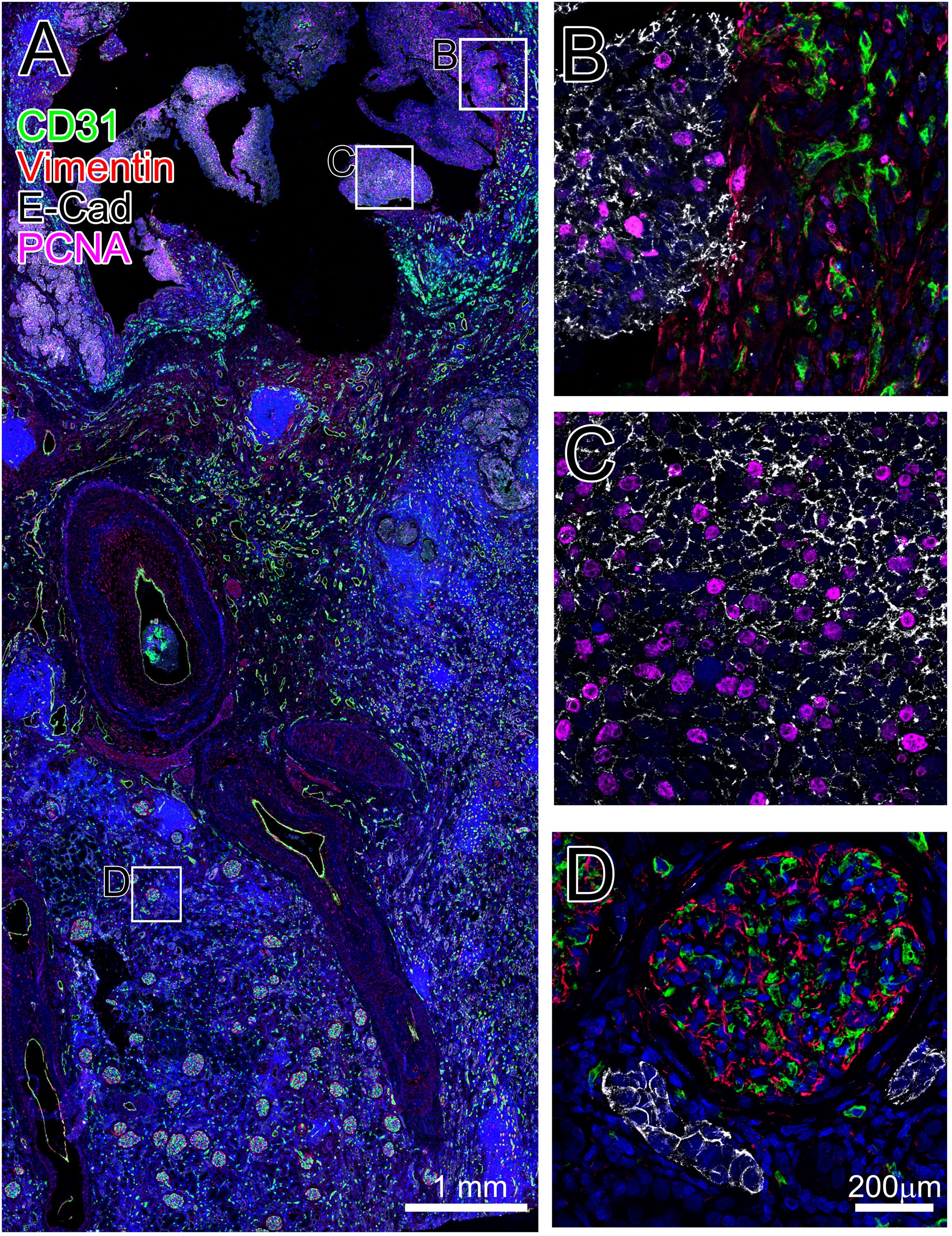
Staining results in human renal tumor tissue confirm the super multi-labeling potential of TSA: successful four-color simultaneous fluorescence staining of CD31, Vimentin, E-Cad, and PCNA was achieved, marking four core targets—vascular endothelium, stromal cells, epithelial cells, and proliferating cells respectively. Each marker signal is independent with no overlap or crosstalk, and specificity is extremely strong. Multiple key information including tumor proliferation, epithelial-mesenchymal transition, angiogenesis, and tissue structure distribution can be simultaneously analyzed on a single tissue section. Compared with traditional technologies, TSA significantly improves single-sample data utilization and is a core tool for tumor multi-omics and microenvironment research.
5. Breaking Species Constraints: Supporting Multi-Staining with Same-Species Antibodies, Completely Liberating Antibody Selection
Traditional IF/IHC multi-labeling has a strict rule: same-species primary antibodies are strictly prohibited from being used together. Multiple homologous antibodies combined will cause severe cross-reactions and whole-slide background contamination, so experiments must forcibly use different-species antibodies. However, many highly specific, gold-standard antibodies only come from a single species source, greatly limiting experimental design. Many studies cannot be conducted due to the lack of suitable heterologous antibodies.
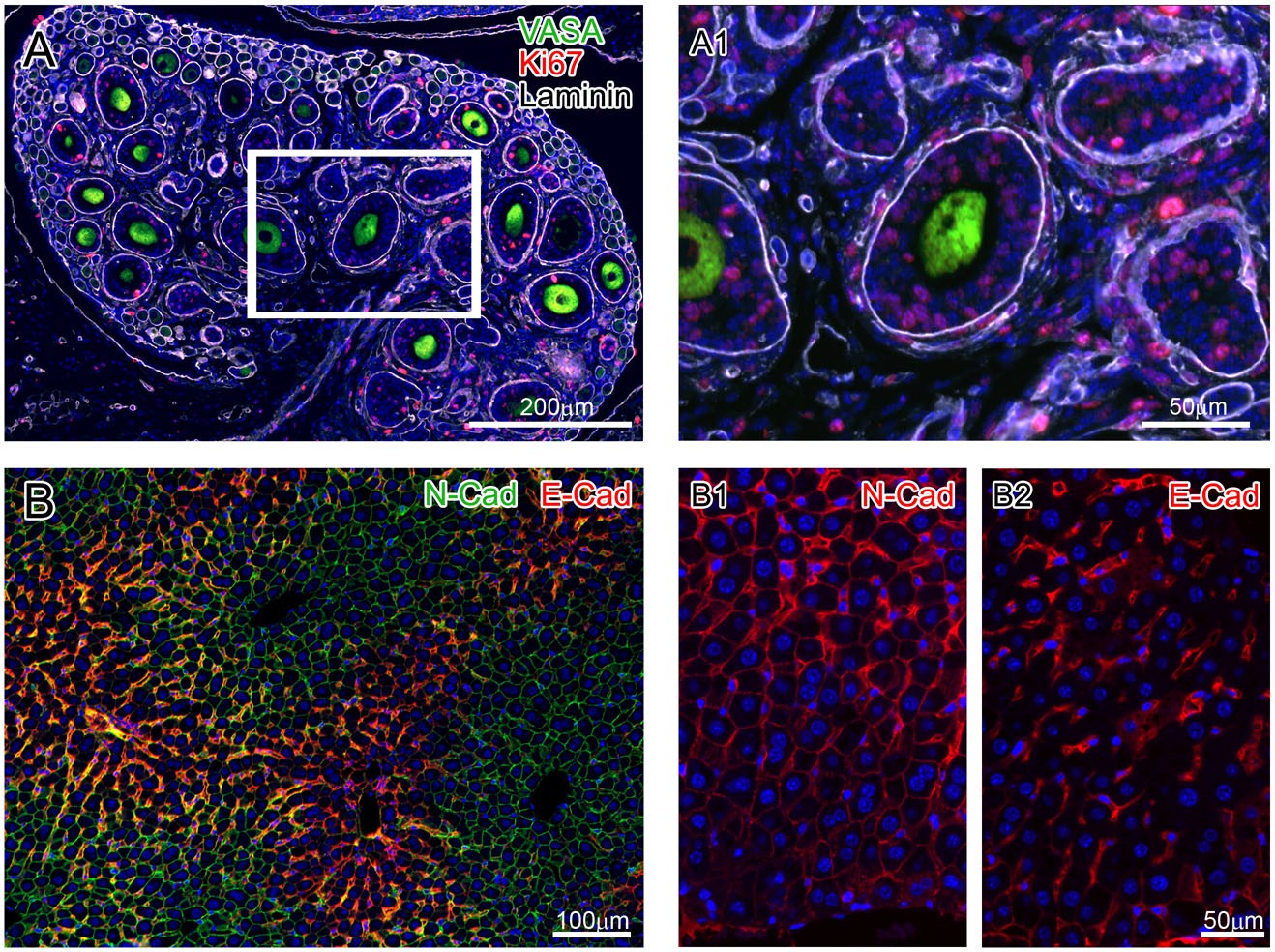
In contrast, TSA's unique site saturation blocking and step-by-step iterative staining technology completely breaks this industry barrier. Multiple control experiments successfully achieved two sets of classic same-species antibody multi-labeling: three rabbit-derived antibodies (VASA, Ki67, Laminin) used together in mouse ovarian tissue, and two mouse-derived antibodies (N-Cad, E-Cad) used together in mouse liver tissue. Staining results showed no cross-reactions, no signal interference, precise target localization, and were completely consistent with single-label staining results. This truly achieves "unlimited species, arbitrary combination," completely solving the problem of high-quality antibody compatibility.
6. Universal Adaptability to Multiple Scenarios: Precise Imaging of Multiple Tissues and Targets in Embryonic Development
Traditional staining technologies have extremely poor adaptability to complex embryonic tissues: embryonic tissue structures are delicate, antigens are easily lost, and cells are dense. Conventional staining easily produces problems such as messy signals, target loss, and excessive non-specific background, making it difficult to achieve simultaneous analysis of multiple structures and targets, which severely limits developmental biology research.
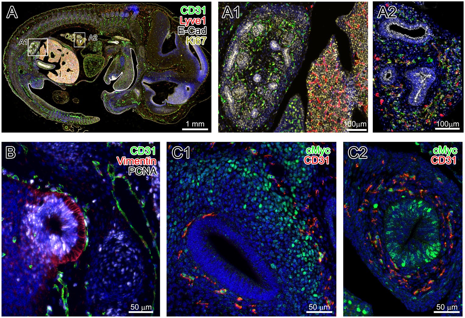
Multi-color staining results of E13.5 mouse embryos comprehensively confirm the broad adaptability of TSA: on the same embryonic paraffin section, simultaneous staining of multiple target types and tissue structures—including vascular endothelium, lymphatic vessels, epithelial cells, proliferating cells, and stromal cells—can be completed, clearly outlining the fine structures and cell proliferation and differentiation characteristics of different organs such as embryonic liver, kidney, lung, cochlea, and midgut. Whether for multi-species, multi-tissue, or complex embryonic samples, TSA can stably output high-definition, specific, and quantifiable staining results, with far superior versatility and stability compared to traditional IHC/IF.
Summary: 6 Experimental Evidences Demonstrate TSA's Comprehensive Overwhelm of Traditional Staining Technologies
Combined with 6 groups of core control experiments, we can clearly summarize the irreplaceable core value of TSA, with each item supported by experimental data:
| Comparison Dimension | Core Advantages of TSA Technology | Limitations of Traditional IHC/IF |
|---|---|---|
| Sensitivity | Ultra-high sensitivity, stably detects low-abundance weakly expressed targets, no false negatives | Only high-expression targets visible, low-abundance signals lost, prone to false negatives |
| Multi-label Typing Capability | Multi-color without crosstalk, clearly distinguishes immune subsets, precisely validates co-localization | Signal overlap with blurred boundaries, unable to perform fine cell typing |
| Anti-Nonspecific Background | Inhibits plasma cell endogenous Ig false staining, clean background for easy interpretation | Endogenous immunoglobulins cause massive false positives, interfering with results |
| Multi-label Throughput | Stably achieves four-color labeling on a single section, multi-dimensional simultaneous analysis of tissue characteristics | Maximum dual-color labeling, severe crosstalk with three or more colors, requires multiple sections for splicing |
| Antibody Species Compatibility | Same-species primary antibodies can be freely combined, no cross-reactions | Limited to different-species antibodies, high-quality homologous antibodies cannot be used |
| Sample Adaptation Scenarios | Stable imaging across embryonic, tumor, and immune tissue scenarios | Complex embryonic samples have messy signals, antigen loss, and high background |
Compared with the numerous limitations of traditional IHC/IF, TSA technology achieves comprehensive upgrades across six dimensions: sensitivity, specificity, compatibility, throughput, adaptability, and precision. Results are stable, reproducible, and precisely quantifiable, making TSA the standard technology for high-impact pathology, immunology, and developmental research.
As the current core technology for multiplex immunofluorescence and high-sensitivity tissue detection, TSA perfectly adapts to various high-end research scenarios including tumor mechanism research, immune microenvironment analysis, embryonic development research, and clinical pathology biomarker validation. It is an essential technology for breaking through traditional experimental bottlenecks, improving research data quality, and facilitating the publication of high-impact papers!
EnkiLife not only provides customers with a complete set of TSA multiplex labeling kits, but also offers various TSA specialty technical services, including IF fluorescence staining, fluorescence panoramic scanning, ultra-multiplex staining, and pathological analysis (5 markers and below).
Product | Catalog Number |
|---|---|
TSA Six-Label Seven-Color Multiplex Immunohistochemistry Kit | |
TSA Five-Label Six-Color Multiplex Immunohistochemistry Kit | |
TSA Four-Label Five-Color Multiplex Immunohistochemistry Kit | |
TSA Three-Label Four-Color Multiplex Immunohistochemistry Kit | |
TSA Two-Label Three-Color Multiplex Immunohistochemistry Kit |
References
Yarilin D, Xu K, Turkekul M, Fan N, Romin Y, Fijisawa S, Barlas A, Manova-Todorova K. Machine-based method for multiplex in situ molecular characterization of tissues by immunofluorescence detection. Sci Rep. 2015 Mar 31; 5:9534. doi: 10.1038/srep09534. PMID: 25826597; PMCID: PMC4821037.