
Pericyte Markers
The BBB is formed by microvascular endothelial cells (ECs) lining the cerebral capillaries penetrating the brain and spinal cord of most mammals, providing a biological barrier at the blood to brain interface effectively separating the brain from the rest of the body (1). Pericytes are essential constituents of the brain capillary; their close association with ECs allows the exchange of ions, metabolites, second messengers, and ribonucleic acids between the two cell types. Pericyte markers include smooth muscle actin, desmin, or platelet-derived growth factor receptor β (PDGFR-β). Pericytes and ECs are in close communication with each other, for example, via the PDGF-β signaling pathway.
The cytoskeleton consists of three types of cytosolic fibers: microfilaments (actin filaments), intermediate filaments, and microtubules. Actin proteins are major components of the eukaryotic cytoskeleton. At least six vertebrate actin isoforms have been identified. The cytoplasmic β- and γ-actin proteins are referred to as “non-muscle” actin proteins as they are predominantly expressed in non-muscle cells where they control cell structure and motility. The α-cardiac and α-skeletal actin proteins are expressed in striated cardiac and skeletal muscles, respectively. The smooth muscle α-actin and γ-actin proteins are found primarily in vascular smooth muscle and enteric smooth muscle, respectively. The α-smooth muscle actin (ACTA2) is also known as aortic smooth muscle actin. These actin isoforms regulate the contractile potential of muscle cells.
The intermediate filament desmin is a myogenic marker expressed in early development that forms a network of filaments extending across the myofibril and surrounding Z discs. The desmin cytoskeleton provides a connection among myofibrils, organelles, and the cytoskeleton . Melanoma cell adhesion molecule (MCAM, MUC18, CD146) is a marker protein seen in vascular endothelial cells, activated T lymphocytes, smooth muscle, and bone marrow stromal cells. Endothelial MCAM within the BBB acts as adhesion receptors that permit lymphocytes to transmigrate across the barrier and produce inflammatory lesions. MCAM also functions as a co-receptor for PDGFR-β on pericytes to regulate pericyte-EC interactions.
CD31 (Platelet Endothelial Cell Adhesion Molecule-1: PECAM-1), a member of the Ig superfamily of cell adhesion molecules, is expressed by circulating platelets, monocytes, neutrophils, some T cells, and endothelial cells and modulates cell adhesion, endothelial cell migration, and angiogenesis. CD31 serves as a scaffold for various signaling molecules and is a possible gate-keeper receptor in the inflammatory blood–brain axis.
Aminopeptidase N (APN, CD13) is a metalloprotease expressed on myeloid cells, pericytes, fibroblasts, epithelial and endothelial cells, as well as on tumor cells, and stem cells. APN/CD13 acts as an adhesion molecule that modulates inflammatory immune cell trafficking, resulting in injury progression.
The chondroitin sulfate proteoglycan NG2 is a type I membrane protein expressed by subpopulations of glia, including oligodendroglial precursor cells and a variety of tumor cells. Although NG2 is not expressed in adult central nervous system (CNS) pericytes, it is an early marker of pericyte activation during CNS development and pathological conditions. Thus, upon CNS injury, NG2-reactive pericytes are found along microvessels, where they act as sensors for inflammation and support the immunosurveillance and effector function of extravasated neutrophils and macrophages.
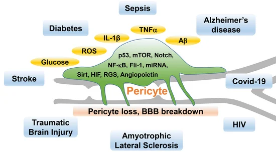
Schematic Diagram of the Mechanisms Linking Pericyte Function to Disease
Relevent antibodies
| Catalog# | Product Name | Reactivity | Application |
|---|---|---|---|
| AMRe21274 | PDGFR-β Rabbit Monoclonal antibody | Human,Mouse,Rat | WB,IHC,IF,IP,ELISA |
| AMRe21513 | α smooth muscle actin Rabbit Monoclonal antibody | Human,Mouse,Rat | WB,IHC,IF,IP,ELISA |
| AMM00779 | Desmin (4H1) Mouse Monoclonal Antibody | Human,Rat,Mouse | IHC-P |
| AMRe21362 | CD146 Rabbit Monoclonal antibody | Human,Mouse,Rat | WB,IHC,IF,IP,ELISA |
| AMRe08200 | CD13 (14U5) Rabbit Monoclonal Antibody | Human,Rat | WB,IHC-P,ICC/IF,IP,IF-P |
| AMRe21591 | CD31 Rabbit Monoclonal antibody | Human, | WB,IHC,IF,IP,ELISA |
| AMM03503 | CD31 (2D3) Mouse Monoclonal Antibody | Human | WB,IP |
| AMRe14671 | NG2 (18P16) Rabbit Monoclonal Antibody | Human | WB,IHC-P,ICC/IF,IF-P |
| APS0631 | HRP-conjugated Polyclonal Goat Anti-Mouse IgG(H+L) Secondary Antibody | ||
| APS0635 | HRP-conjugated Polyclonal Goat Anti-Rabbit IgG(H+L) Secondary Antibody | ||
| AMre80004 | GAPDH (12R9) Rabbit Monoclonal Antibody |
Related Products
References
- Kadry H, Noorani B, Cucullo L. A blood-brain barrier overview on structure, function, impairment, and biomarkers of integrity. Fluids Barriers CNS. 2020 Nov 18;17(1):69.[PMID: 33208141].
- Eng LF, Ghirnikar RS, Lee YL. Glial fibrillary acidic protein: GFAP-thirty-one years (1969-2000). Neurochem Res. 2000 Oct;25(9-10):1439-51. doi: 10.1023/a:1007677003387. PMID: 11059815.
- Zhang Z, Gan Q, Han J, Tao Q, Qiu WQ, Madri JA. CD31 as a probable responding and gate-keeping protein of the blood-brain barrier and the risk of Alzheimer's disease. J Cereb Blood Flow Metab. 2023 Jul;43(7):1027-1041.[PMID: 37051650].
- Li P, Fan H. Pericyte Loss in Diseases. Cells. 2023 Jul 26;12(15):1931. [PMID: 3756601].
 | Flora Flora is a technical support expert at EnkiLife, familiar with immunology and neuroscience, dedicated to providing customers with high-quality product combinations and technical support to help achieve research in neurodegenerative diseases and other neuroscience areas. |