
The tumor immune microenvironment (TIME) is a complex dynamic microecosystem composed of tumor cells, infiltrating immune cells, stromal cells, cytokines, and extracellular matrix. Its cellular components, spatial distribution, and interaction patterns directly determine tumor proliferation, invasion, metastasis, and immunotherapy response. Traditional single immunohistochemistry and single-color immunofluorescence staining can only detect a single biomarker, failing to restore the multi-cellular and multi-molecular regulatory characteristics of TIME, with obvious detection limitations. mIHC multiplex immunostaining technology can simultaneously localize and quantify more than ten or even dozens of protein markers on a single tissue section, accurately analyzing the infiltration density, spatial arrangement, functional status, and intercellular interactions of various immune cells. It has been widely applied in cutting-edge research such as tumor microenvironment, spatial proteomics, neuroscience, and pathological classification, becoming a core supporting technology for high-impact journals. This article systematically elaborates the specific application scenarios and clinical value of multiplex immunostaining technology in tumor immune microenvironment research, combined with multiple research examples.
Significant immune microenvironment heterogeneity exists between the tumor nest and stromal regions of non-small cell lung cancer (NSCLC). This heterogeneity directly shapes the immunosuppressive microenvironment and is a key inducer of lung cancer immune escape and disease progression. The expression and spatial distribution of immune markers are the core basis for determining the degree of TIME suppression.
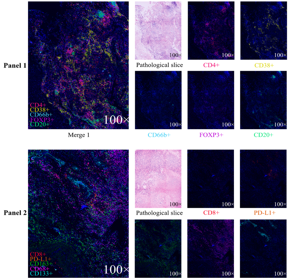
The Peng team used mIHC multiplex immunofluorescence technology to analyze the expression characteristics and cellular interaction relationships of 10 core immune markers in the tumor nest and tumor stroma regions of NSCLC.
The results clearly confirmed the existence of multiple key immune cell interaction systems in the lung cancer microenvironment: CD20⁺ B cells are strongly associated with CD4⁺CD38⁺ T cells, and neutrophils are correlated with FOXP3⁺ regulatory T cells in the stromal region; the interactions between CD8⁺ T cells and PD-L1⁺ M2 macrophages, as well as between PD-L1-positive cells and CD8⁺ T cells within the tumor, directly affect anti-tumor immune responses. The study also found a moderate correlation between tumor CD133⁺ stem cell-like cells and non-PD-L1-expressing M1 macrophages, suggesting that macrophages may participate in mediating CD8⁺ T cell exhaustion.
This study, leveraging the multi-target simultaneous detection advantage of mIHC, for the first time completely outlined the tumor immune landscape and cellular interaction network of NSCLC, providing critical data support for in-depth lung cancer mechanism research and new therapeutic target screening.
Clinical tumor research often requires batch analysis of samples. Traditional staining techniques are cumbersome and inefficient, making it difficult to meet high-throughput research needs. The combined approach of mIHC multiplex immunofluorescence + tissue microarray (TMA) perfectly solves this problem.
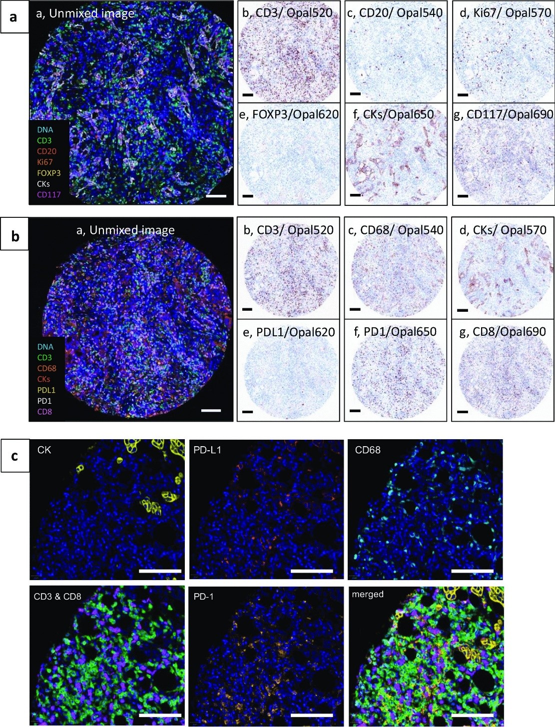
This study optimized two standardized multiplex immunofluorescence staining panels, IP1 and IP2, for high-throughput multi-marker simultaneous labeling on bronchial lung cancer and breast cancer tissue microarrays. Images were collected using the Vectra 3 imaging system, followed by multispectral image unmixing and pseudo-color assignment with inForm software. It can also generate simulated DAB-stained pathological views, balancing the accuracy of multi-index simultaneous detection with conventional pathological interpretation habits.
The IP1 panel can simultaneously identify 5 core cell types in the TME, accurately assess the proliferative activity of various cells combined with Ki67 nuclear staining, and precisely distinguish tumor cells and mast cell subsets through CK and CD117 protein co-localization. The IP2 panel focuses on core immune cells such as conventional T cells, cytotoxic T cells, and macrophages, while including PD-1/PD-L1 immune checkpoint detection, successfully capturing the key feature of PD-L1⁺CD68⁺ macrophages colocalizing tightly with PD-1⁺ T cells in situ in breast cancer samples.
This study validated the advantages of mIF combined with TMA technology, enabling systematic in-situ analysis of microenvironment cell typing, proliferative functional status, and immune cell spatial molecular interactions across multiple tumor types on a single tissue section. It provides a standardized, implementable technical pathway for large-scale clinical sample tumor immune microenvironment visualization research.
The development and progression of colorectal cancer are closely related to the abnormal expression of multiple protein markers. Precise localization and quantitative detection of various markers are important foundations for tumor classification and prognosis prediction.
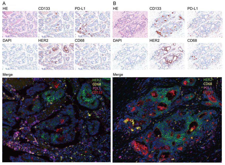
Zhang et al. conducted mIHC multiplex staining experiments on colorectal cancer tissues, performing multi-target detection on sigmoid colon cancer and ascending colon cancer tissues respectively. The experiment used HE staining and DAPI staining for tissue localization, followed by detection of the expression distribution of four core proteins: CD133, PD-L1, HER2, and CD68.
The results showed that the four proteins have different expression locations and functions in colorectal cancer tissues: CD133 is enriched on the surface of cancer cells and is associated with tumor stem cell characteristics; PD-L1 is expressed on cancer cells and immune cells, mediating immune suppression; HER2 is localized on the cancer cell membrane and is a core biomarker for targeted therapy; CD68 specifically marks macrophages in the tumor microenvironment.
Compared with traditional single-label staining, mIHC multi-color fusion images can clearly distinguish the spatial distribution and co-expression characteristics of the four proteins on the same section, with no spectral interference and precise signals. It provides reliable technical support for colorectal cancer marker research, precise classification, and efficacy prediction.
Tertiary lymphoid structures (TLS) are key immune functional structures in the tumor microenvironment, closely related to tumor immune responses and immunotherapy efficacy. However, their spatial distribution, cellular composition, and functional characteristics in gliomas have long lacked precise analysis methods.
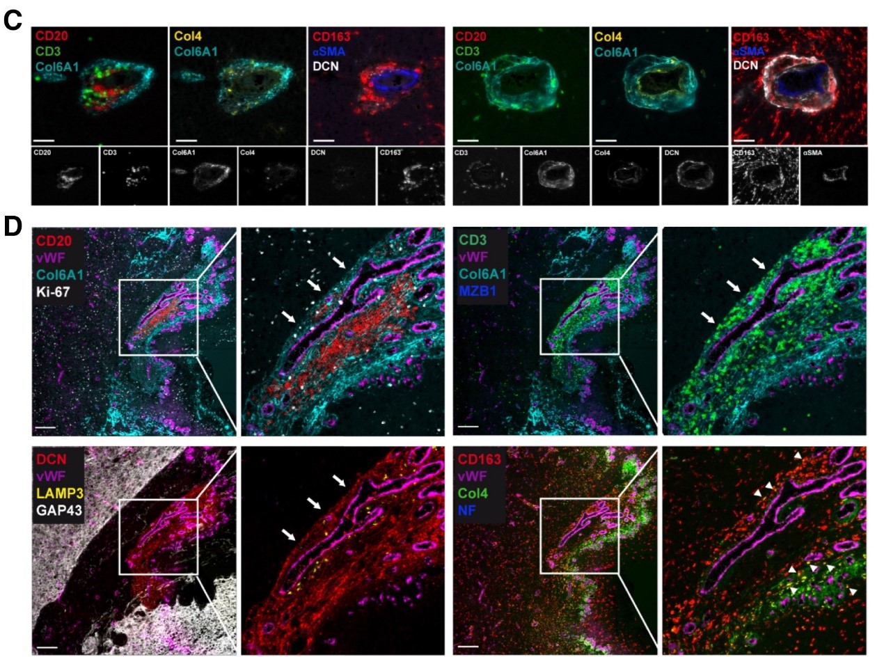
The Cakmak team used multi-marker combination mIHC technology to precisely analyze the spatial structure and cellular components of functional TLS in gliomas. The study clearly demonstrated the distribution patterns of CD20⁺ B cells and CD3⁺ T cells in the perivascular microenvironment, while simultaneously localizing the expression positions of extracellular matrix components such as Col6A1 and Col4, and functional proteins such as αSMA and CD163, clarifying the spatial association between immune cells and stromal components.
Key findings: Glioma TLS aggregates in Col6A1-positive dense extracellular matrix, adjacent to vascular tissue; TLS contains various functional immune cells including proliferating cells, activated B cells, and dendritic cells, with complete immune response capabilities. It was also confirmed that CD163⁺Col4⁺ double-positive macrophages can uptake stromal components, and this functional TLS is specifically distributed within the tumor, with Col6A1 expressed only in tumor areas and absent in non-tumor areas.
Relying on the high-precision spatial analysis capability of mIHC, this study for the first time clarified the characteristics of functional TLS subsets in gliomas, opening new directions for glioma immune classification and immune mechanism research.
Triple-negative breast cancer (TNBC) has high malignancy and large prognostic differences. Precise screening of prognosis-related immune markers is crucial for clinical treatment decision-making.
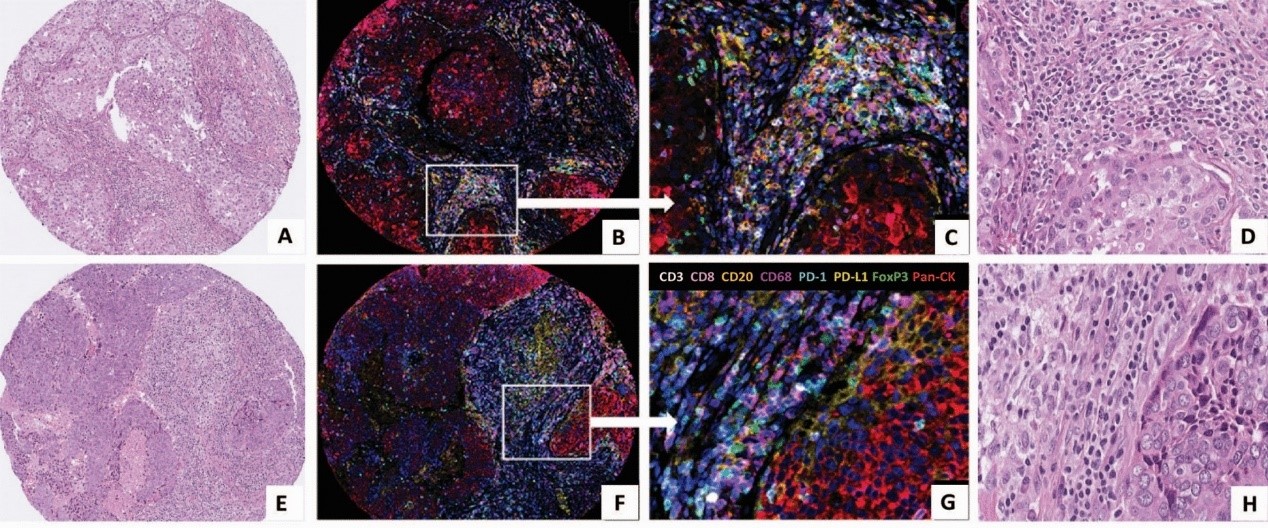
The Wang team used 8-color mIHC multiplex immunofluorescence technology to detect tumor tissue sections from TNBC patients. The study first used H&E staining to clarify the macroscopic morphology of tumor tissue and stromal regions, providing pathological background for immune cell localization. Subsequently, relying on a high-resolution multi-color fluorescence system, various immune cell marker signals were precisely distinguished without cross-interference.
The experiment successfully identified the key cell subset of high stromal CD68⁺PD-L1⁺ macrophages: target cells were precisely identified through CD68 and PD-L1 signal co-localization, while Pan-CK signals were used to exclude tumor cell interference, ensuring the accuracy and reliability of detection results. Subsequent clinical data analysis confirmed that this specific macrophage subset can serve as an independent predictor of improved prognosis in triple-negative breast cancer patients, providing a novel biomarker for breast cancer prognosis assessment and immunotherapy efficacy prediction.
From lung cancer, breast cancer, colorectal cancer to glioma, numerous high-impact studies have fully confirmed: mIHC multiplex immunostaining, with its core advantages of multi-target detection on single sections, precise spatial localization, quantitative analysis capability, and adaptability to high-throughput samples, perfectly compensates for the shortcomings of traditional staining techniques. It can deeply analyze the cellular interactions, marker expressions, and spatial structural characteristics of the tumor immune microenvironment. It is currently the core technology for tumor mechanism research, biomarker screening, prognosis analysis, and immunotherapy research, and is also a key driver for publishing high-impact SCI papers.
Professional mIHC multiplex immunofluorescence TSA technical services from EnkiLife provide one-stop coverage of the entire experimental workflow for tumor microenvironment research. With standardized antibody ratios, spectral unmixing, and spatial quantitative analysis, it avoids common problems such as signal interference and uneven staining in self-conducted experiments, efficiently supporting the production of high-impact SCI research outcomes.
Product | Catalog Number |
|---|---|
TSA Six-Label Seven-Color Multiplex Immunohistochemistry Kit | |
TSA Five-Label Six-Color Multiplex Immunohistochemistry Kit | |
TSA Four-Label Five-Color Multiplex Immunohistochemistry Kit | |
TSA Three-Label Four-Color Multiplex Immunohistochemistry Kit | |
TSA Two-Label Three-Color Multiplex Immunohistochemistry Kit |
References
[1] Peng H, Wu X, Zhong R, Yu T, Cai X, Liu J, Wen Y, Ao Y, Chen J, Li Y, He M, Li C, Zheng H, Chen Y, Pan Z, He J, Liang W. Profiling Tumor Immune Microenvironment of Non-Small Cell Lung Cancer Using Multiplex Immunofluorescence. Front Immunol. 2021 Nov 4;12:750046. doi: 10.3389/fimmu.2021.750046. PMID: 34804034; PMCID: PMC8600321.
[2] Mori H, Bolen J, Schuetter L, Massion P, Hoyt CC, VandenBerg S, Esserman L, Borowsky AD, Campbell MJ. Characterizing the Tumor Immune Microenvironment with Tyramide-Based Multiplex Immunofluorescence. J Mammary Gland Biol Neoplasia. 2020 Dec;25(4):417-432. doi: 10.1007/s10911-021-09479-2. Epub 2021 Feb 15. PMID: 33590360; PMCID: PMC7960613.
[3] Zhang W, Song ZJ, Zhang BY, Wang JL, Guo Q, Sun ZW, Tang H. Multiplex immunohistochemistry indicates biomarkers in colorectal cancer. Neoplasma. 2021 Nov;68(6):1272-1282. doi: 10.4149/neo_2021_210312N324. Epub 2021 Aug 30. PMID: 34459208.
[4] Cakmak P, Lun JH, Singh A, Macas J, Schupp J, Schuck J, Mahmoud Z, Köhler M, Starzetz T, Burger MC, Steidl E, Hasse LM, Hattingen E, Plate KH, Reiss Y, Imkeller K. Spatial immune profiling defines a subset of human gliomas with functional tertiary lymphoid structures. Immunity. 2025 Nov 11;58(11):2847-2863.e8. doi: 10.1016/j.immuni.2025.09.018. Epub 2025 Oct 21. PMID: 41125076.
[5] Wang J, Browne L, Slapetova I, Shang F, Lee K, Lynch J, Beretov J, Whan R, Graham PH, Millar EKA. Multiplexed immunofluorescence identifies high stromal CD68+PD-L1+ macrophages as a predictor of improved survival in triple negative breast cancer. Sci Rep. 2021 Nov 3;11(1):21608. doi: 10.1038/s41598-021-01116-6. PMID: 34732817; PMCID: PMC8566595.