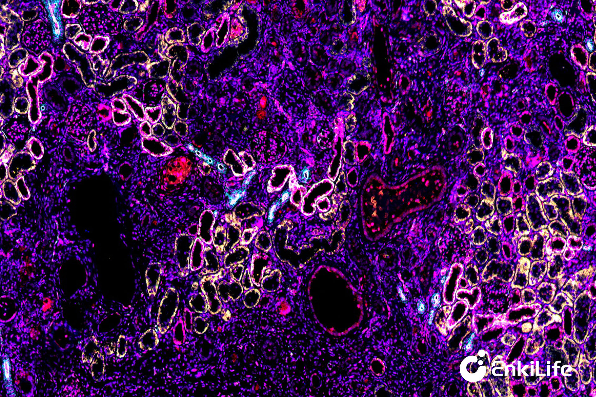
Many researchers have fallen into the same misconception: as long as the sections are successfully stained, usable research results can be produced. But real mIHC experiments are never "finished after staining." From an ordinary tissue sample to a complete, precise, and publishable data system, every step requires strict quality control. Any oversight in any link will cause staining distortion, data deviation, and spatial information failure, ultimately rendering the entire experiment useless and the paper data unusable.
This article will guide you through the core steps of an mIHC project from raw sections to top-tier journal figures, helping you thoroughly understand the research logic and application value of mIHC.
Basic Sample Preparation: Standardized processing of FFPE paraffin tissues and frozen tissues to ensure complete tissue morphology and intact antigen preservation.
Section Scheme Design: Planning serial sections or tissue microarrays (TMA) according to research needs to avoid sample bias.
Pre-Quality Verification: Screening for section wrinkles, detachment, and impurities, confirming intact antigen preservation in tissues.
Custom Panel Design: Customizing marker combinations based on research targets.
Whole-slide panoramic imaging with no field-of-view omissions and no edge artifacts
Multi-channel fluorescence simultaneous acquisition with precise matching of each target's fluorescence wavelength band
High-resolution digital archiving preserving extreme detail for subsequent refined analysis
| Data Category | Detection Content |
|---|---|
| Basic Quantification | Precise cell identification, positive cell counting, positive rate statistics, fluorescence expression intensity analysis |
| Core Spatial Analysis | Cell co-localization determination, intercellular spatial distance measurement, immune cell aggregation characteristic analysis |