
Herpesviridae refers to a viral family consisting of enveloped double‑stranded DNA viruses with large, sophisticated genomes, generally called herpesviruses. These pathogens replicate inside the cell nucleus of numerous vertebrate species. Among over a hundred identified herpesvirus strains, nine distinct types commonly infect human bodies, namely herpes simplex virus 1, herpes simplex virus 2, varicella‑zoster virus, Epstein‑Barr virus, cytomegalovirus, human herpesvirus‑6, human herpesvirus‑7, and Kaposi's sarcoma‑associated herpesvirus.
This article focuses mainly on human herpesviruses, covering their viral morphology, taxonomic classification, related clinical disorders and transmission pathways.
The genome of herpesviruses is composed of non‑segmented linear double‑stranded DNA. Its length ranges roughly from 120 to 250 kbp, encoding 70 to 200 distinct viral genes.
Herpesviruses feature a distinctive four‑tier structural layout:
- Core: This innermost compartment stores a single linear torus‑shaped dsDNA molecule.
- Capsid: Wrapping the core is an icosahedral shell with a diameter of 100 nm, assembled from 162 capsomer subunits.
- Tegument: Situated between the capsid and outer envelope lies an unstructured, occasionally asymmetric layer. It contains various viral enzymes: some hijack host cellular metabolic pathways to support viral particle synthesis, others counteract the host’s immediate immune defenses, while the biological functions of several remaining components remain unclarified.
- Envelope: Serving as the outermost layer of viral particles, it is formed by modified host cell membrane embedded with over ten unique viral glycoproteins. Virions appear spherical or polymorphic, measuring 150–200 nm across and carrying T=16 icosahedral symmetry.
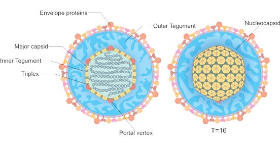
Figure 1. Structural schematic of herpesvirus particles
Herpesviruses fall into three separate subfamilies based on biological traits:
- Alpha herpesviruses: Comprising HSV‑1, HSV‑2 and varicella‑zoster virus. They exhibit fast replication cycles, trigger cytopathic effects in monolayer cell cultures and possess a broad spectrum of susceptible hosts.
- Beta herpesviruses: Including cytomegalovirus, human herpesvirus 6 and human herpesvirus 7. Their replication cycle is prolonged, and their host tropism is relatively limited.
- Gamma herpesviruses: Covering Epstein‑Barr virus and human herpesvirus 8, which display extremely narrow host specificity.
At present, more than 130 herpesvirus variants have been discovered, isolated from mammals, birds, fish, reptiles, amphibians and mollusks. Nine subtypes are confirmed to infect human beings.
Table 1: Taxonomic grouping of human herpesviruses
| Name | Genus | Alternative Name | Subfamily | Primary Target Cell |
|---|---|---|---|---|
| HHV‑1 | Simplexvirus | Herpes simplex virus type 1 (HSV‑1) | Alpha (α) | Mucoepithelial cells |
| HHV‑2 | Simplexvirus | Herpes simplex virus type 2 (HSV‑2) | Alpha (α) | Mucoepithelial cells |
| HHV‑3 | Varicellovirus | Varicella zoster virus (VZV) | Alpha (α) | Mucoepithelial cells |
| HHV‑5 | Lymphocryptovirus | Cytomegalovirus (CMV) | Beta (β) | Monocytes and epithelial cells |
| HHV‑6A and 6B | Roseolovirus | Human herpesvirus 6 | Beta (β) | T lymphocytes |
| HHV‑7 | Roseolovirus | Human herpesvirus 7 | Beta (β) | T lymphocytes |
| HHV‑4 | Lymphocryptovirus | Epstein–Barr virus (EBV) | Gamma (γ) | B cells and epithelial cells |
| HHV‑8 | Rhadinovirus | Kaposi's Sarcoma‑associated herpesvirus (KSHV) | Gamma (γ) | Lymphocytes and other somatic cells |
The table below summarizes illnesses triggered by human herpesviruses alongside their typical clinical manifestations.
Table 2: Clinical diseases associated with human herpesviruses
| Virus | Corresponding Diseases | Typical Signs & Symptoms |
|---|---|---|
| HSV1, HSV2 | Herpes simplex infection | Skin blisters, mucosal ulcers, oral fever blisters and general flu‑like malaise |
| VZV | Chickenpox, Herpes zoster (shingles) | Generalized skin rash, fluid‑filled blisters, fever, neuralgia, sore throat, headache and abdominal discomfort |
| CMV | Cytomegalovirus infection, CMV mononucleosis | Fever, cutaneous rash, pharyngalgia, nausea, myalgia, swollen lymph nodes and persistent fatigue |
| EBV | EBV‑induced infectious mononucleosis; linked to Burkitt’s lymphoma and multiple malignant tumors | Fever, rash, sore throat, nausea, muscle soreness, lymphadenectasis, exhaustion, weight loss and vomiting |
| HHV6, HHV7 | Roseola infantum (exanthem subitum) | Hyperthermia, enlarged lymph nodes, rhinorrhea, mild diarrhea, swollen eyelid tissues, fatigue and post‑fever rash |
| KSHV | Kaposi's sarcoma (KS) | Distinctive cutaneous lesions, lymphoma, plus non‑specific symptoms such as fever and weight loss |
Nearly all human herpesviruses spread between individuals via direct physical contact between susceptible populations and infected carriers. A small number of herpesvirus strains can also propagate through respiratory droplets. The transmission modes of human herpesviruses are organized in the following chart.
Table 3: Transmission pathways of human herpesviruses
| Virus | Transmission Mechanisms |
|---|---|
| HHV‑1 (HSV1) | Direct exposure to virus‑containing lesions and bodily secretions |
| HHV‑2 (HSV2) | Sexual contact; vertical transmission during childbirth from mothers with genital herpes lesions |
| HHV‑3 (VZV) | Close physical contact and respiratory aerosol transmission |
| HHV‑4 (EBV) | Saliva exchange, potential sexual transmission, intrauterine vertical transmission |
| HHV‑5 (CMV) | Infectious bodily fluids (urine, saliva), transplacental transmission, organ transplantation and blood transfusion |
| HHV‑8 (KSHV) | Saliva contact and sexual intercourse |