mIHC Multiplex Fluorescence Immunohistochemistry —— TSA Technology
mIHC, short for Multiplex Immunohistochemistry, is a high-throughput, high spatial resolution pathological detection technology iteratively upgraded from traditional immunohistochemistry (IHC). This technology can simultaneously detect the expression levels, localization distribution, and co-expression relationships of multiple protein markers on the same tissue section sample. It can completely preserve the spatial structural information of the tissue microenvironment, accurately analyze cellular heterogeneity, cell-cell interactions, and in situ protein expression characteristics. It is widely used in tumor immunology, targeted drug development, disease mechanism research, clinical pathological diagnosis, and other fields.
I. Introduction to mIHC Technology
mIHC multiplex fluorescence immunohistochemistry is a multiplex detection technology developed to address the pain points of traditional IHC. Its core breakthrough is the simultaneous detection of multiple proteins on a single tissue section. Combined with tyramide signal amplification technology, multi-color fluorescence labeling, spectral imaging, and splitting technology, it has achieved the upgrade of pathological detection from "single-dimensional single indicator" to "multi-dimensional panoramic analysis".
Different from the enzymatic color development of traditional IHC, mIHC uses specific fluorescent groups as signal markers. Through the process of cyclic staining, signal amplification, spectral acquisition, and splitting, it can complete the simultaneous detection of 4-10 or even more protein markers on the same sample. This technology completely preserves the in situ spatial structure of the tissue, can accurately locate the expression position of each protein within cells, simultaneously quantify protein expression intensity and positive cell ratio, and can also analyze the spatial distribution, interaction, and co-expression characteristics of different cells.
Compared with traditional technologies, mIHC has the core advantages of high sample utilization, high detection throughput, strong spatial resolution, precise quantification, and good result repeatability. It is perfectly suitable for high-end scientific research and clinical application scenarios such as tumor immune microenvironment analysis, biomarker screening, drug mechanism research, and clinical prognosis assessment.
II. TSA Technology Principle
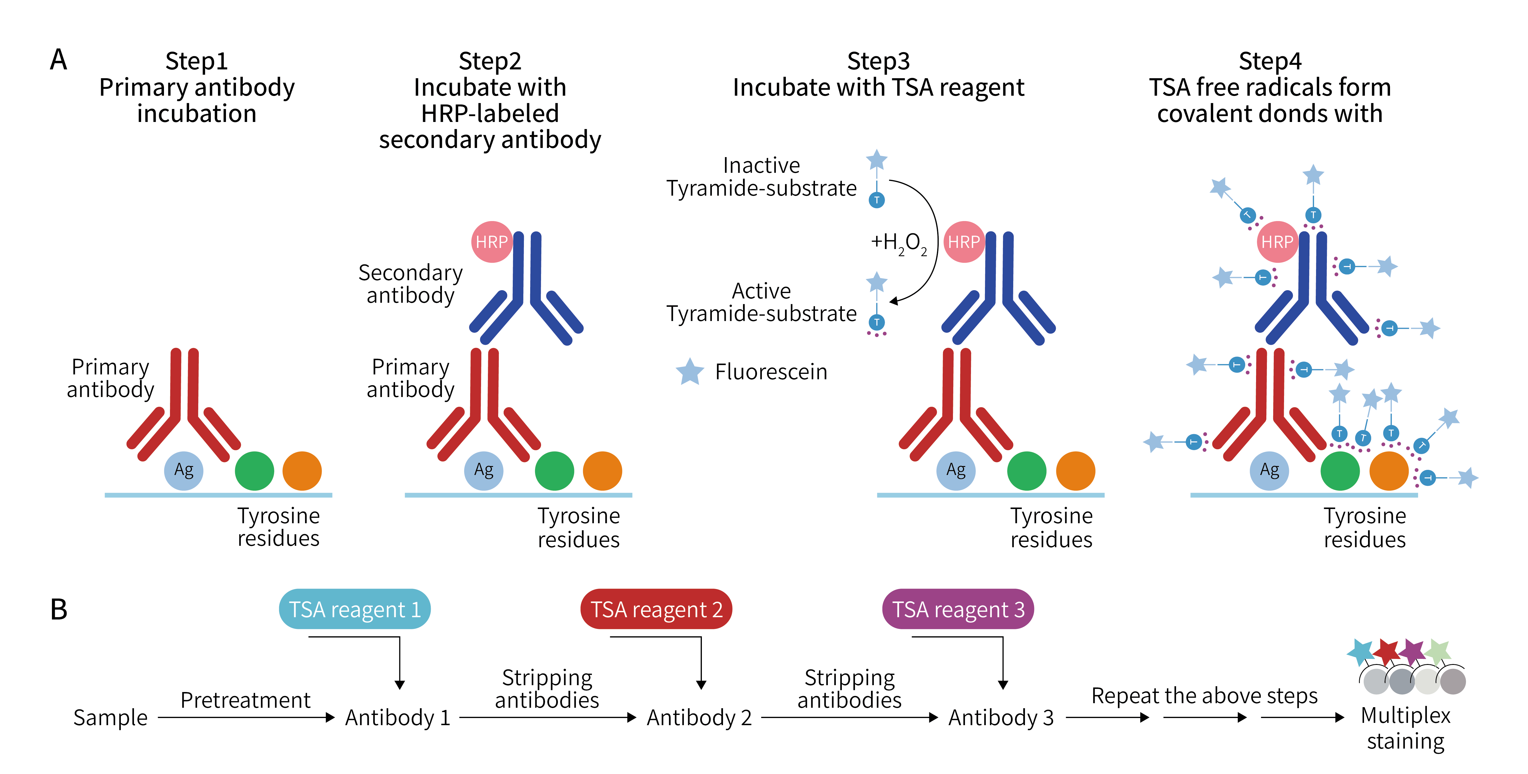
TSA (Tyramide Signal Amplification) is the core underlying technology that enables mIHC to achieve high sensitivity and multi-marker co-staining. It completely solves the technical bottlenecks of weak signals and inability to perform multiplex staining in traditional IHC. Its core principle is based on enzyme-catalyzed in situ covalent binding reaction. During the experiment, specific primary antibodies are first used to precisely target and bind to the target protein antigens in the tissue section, forming antigen-primary antibody complexes to ensure detection specificity. Subsequently, secondary antibodies labeled with horseradish peroxidase (HRP) are added, allowing the secondary antibodies to specifically bind to the primary antibodies, thereby anchoring HRP enzyme molecules at the target antigen sites. On this basis, tyramide substrates with fluorescent labels are added. Under the combined action of HRP enzyme and hydrogen peroxide in the system, the tyramide substrate is activated and transformed into highly reactive free radical intermediates. The activated fluorescent tyramide free radicals are extremely unstable and can rapidly undergo irreversible covalent binding with tyrosine residues in tissue proteins around the antigen sites. This causes a large number of fluorescent tyramide molecules to aggregate and deposit in situ at the target sites, ultimately achieving exponential amplification of the detection signal. After signal deposition for the first protein indicator is completed, the primary antibody-secondary antibody complexes in the sample can be eluted using mild reagents, leaving only the fluorescent signals stably covalently bound to the tissue. Then, tyramide substrates labeled with different fluorescent markers are used, and the above binding, catalysis, and signal amplification processes are repeated to sequentially complete staining detection of multiple different protein markers on the same section, achieving multi-indicator cyclic detection.
III. TSA-mIHC Standard Experimental Procedure
1. Tissue Section Deparaffinization and Hydration
2. Antigen Retrieval
3. Blocking Endogenous Enzyme
4. Blocking
5. Primary Antibody and HRP Secondary Antibody Incubation
6. TSA Fluorescence Deposition
7. Heat-Induced Antibody Stripping
8. Multiple Rounds of Cyclic Staining
9. DAPI Nuclear Staining and Mounting
10. Multispectral Scanning and Image Analysis
IV. How TSA Achieves High Sensitivity and Multi-Marker Co-Staining
Traditional IHC relies only on single signal output from antigen-antibody binding, with limited signal intensity. In contrast, TSA technology achieves a hundredfold increase in sensitivity through enzyme-catalyzed in situ signal enrichment and amplification. There are two core reasons:
First, exponential signal amplification: A single HRP enzyme molecule can continuously catalyze the in situ deposition of dozens to hundreds of fluorescent tyramide molecules. Compared with the one-to-one signal binding mode of traditional IHC, the signal intensity increases exponentially, making it easy to capture low-abundance and trace-expressed protein markers in tissues and significantly reducing the false negative rate.
Second, extremely stable signal: The binding between tyramide molecules and tissue proteins is an irreversible covalent bond, and the binding product has high stability. It will not fall off during subsequent elution, washing, and incubation processes, maximizing the retention of target signals, avoiding signal loss, and further improving detection sensitivity and accuracy.
Traditional multiplex staining cannot achieve multi-indicator simultaneous detection due to antibody residue and signal interference. However, TSA technology perfectly solves this technical problem through a controllable cyclic staining system:
First, elutable antibody system with no cross-interference. In TSA staining, only fluorescent tyramide forms covalent bonds with the tissue, while primary and secondary antibodies are non-covalently bound. After completing the staining and signal fixation for one marker, the primary and secondary antibodies of this round can be completely removed using a dedicated elution buffer, with no antibody residue or signal cross-interference, providing a pure reaction environment for the staining of the next marker.
Second, multi-color fluorescence spectral distinction. By using tyramide substrates labeled with specific fluorescent groups of different emission wavelengths, combined with the precise splitting technology of spectral imaging systems, multiple fluorescent signals can be effectively distinguished, avoiding spectral overlap interference, and achieving independent identification and quantitative analysis of multiple markers on the same section.
Third, cyclic iteration with no sample loss. The entire staining process does not require repeated sectioning. Multi-indicator detection is completed only through multiple cycles of incubation, elution, and signal deposition, with zero sample loss. At the same time, the detection results of all markers correspond to the same tissue field of view, accurately achieving protein co-localization and cell interaction analysis.
EnkiLife not only provides customers with a complete set of TSA multiplex labeling kits, but also offers various TSA specialty technical services, including IF fluorescence staining, fluorescence panoramic scanning, ultra-multiplex staining, and pathological analysis (5 markers and below).
Product | Catalog Number |
|---|---|
TSA Six-Label Seven-Color Multiplex Immunohistochemistry Kit | |
TSA Five-Label Six-Color Multiplex Immunohistochemistry Kit | |
TSA Four-Label Five-Color Multiplex Immunohistochemistry Kit | |
TSA Three-Label Four-Color Multiplex Immunohistochemistry Kit | |
TSA Two-Label Three-Color Multiplex Immunohistochemistry Kit |
For details, please check TSA mIHC Kit