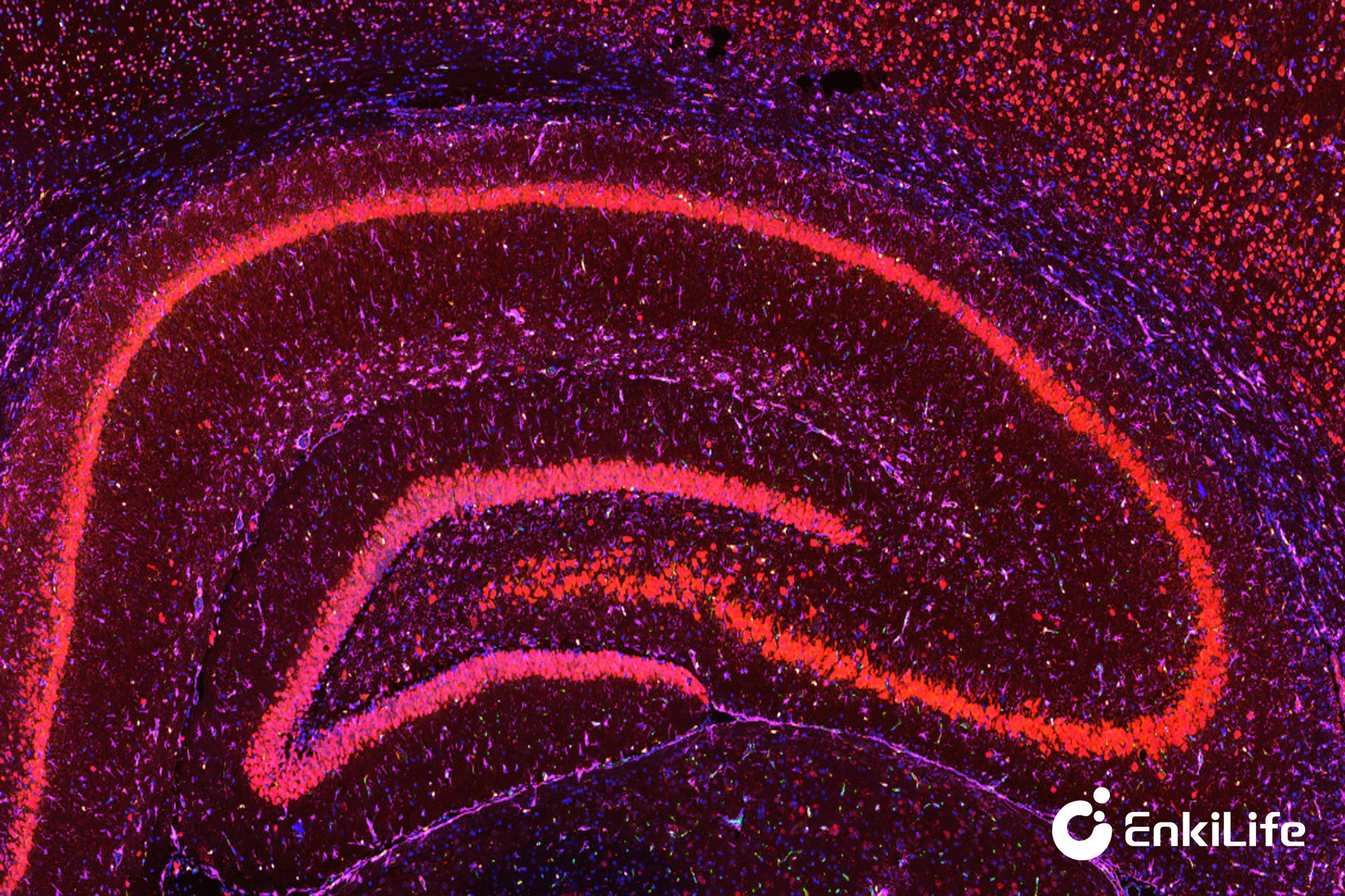
In the fields of life science research and clinical pathological diagnosis, precise identification of trace proteins in tissue samples and realization of simultaneous detection of multiple targets are key to analyzing disease mechanisms and guiding precision medicine. Traditional immunohistochemistry technology is limited by signal intensity and multi-color detection capability, making it difficult to meet the needs of low-abundance target detection and multi-protein co-localization analysis. TSA-mIHC multiplex fluorescence immunohistochemistry technology has become one of the core technologies in current pathological research due to its advantages of ultra-high sensitivity, simultaneous multi-color chromogenesis, and high specificity. Its core competitiveness stems from a precise and unique tyramide signal amplification (TSA) chromogenic reaction system. Today, we will deconstruct the scientific principles of this chromogenic reaction layer by layer, revealing the underlying logic of high-precision pathological staining.
TSA-mIHC stands for Tyramide Signal Amplification-Multiplex Immunohistochemistry, an advanced detection method that integrates TSA tyramide signal amplification technology with traditional multiplex immunohistochemistry technology. Simply put, it is a protein detection technology that relies on enzymatic chemical reactions to achieve signal amplification and combines cyclic staining processes to achieve multi-target chromogenesis.
Traditional immunohistochemistry adopts a "one-to-one" antibody signal binding mode, where one antibody can only carry a small amount of chromogenic signal. For target proteins with extremely low expression levels in tissues, the signal is weak and easily masked by background noise, significantly reducing detection accuracy. The TSA chromogenic reaction completely breaks this limitation by achieving exponential signal amplification through enzyme-catalyzed in situ covalent deposition reactions, while relying on a unique staining cycle mechanism to achieve precise chromogenesis of 7 or more targets on the same tissue section, perfectly adapting to complex tissue microenvironment analysis needs.
All chromogenic effects and signal advantages of TSA-mIHC are built upon three core reactions: horseradish peroxidase (HRP) enzyme-catalyzed reaction, tyramide molecule activation, and in situ covalent deposition. The entire reaction is mild, efficient, and highly specific, which is the key difference from traditional staining techniques.
The core logic of the entire chromogenic system can be summarized as: using antigen-antibody specific binding as the positioning foundation, HRP enzyme as the catalytic core, and fluorescently labeled tyramide as the chromogenic substrate, through a controllable enzyme-catalyzed reaction to generate activated free radicals, achieving stable deposition of large amounts of fluorescent molecules at the target site, ultimately completing signal amplification and specific chromogenesis.
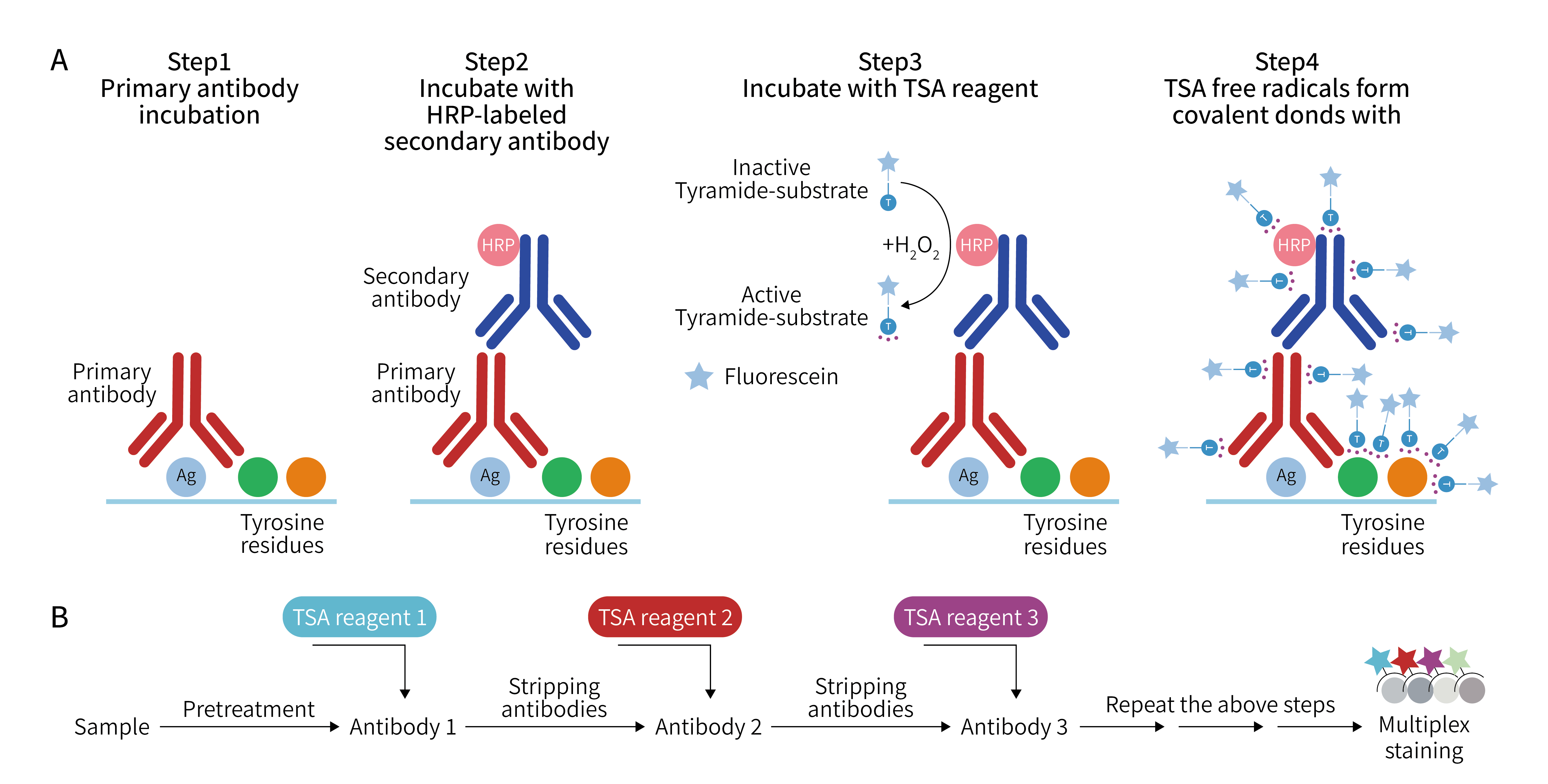
The chromogenic reaction of TSA-mIHC is not a single chemical reaction, but a coherent, precise, and progressive chain reaction. Each step is supported by clear scientific principles, interlocking to ensure the specificity and sensitivity of chromogenesis.
This is the prerequisite for the chromogenic reaction and determines the precision of staining results. In the experiment, specific primary antibodies are first used to incubate tissue sections. The primary antibodies will precisely recognize and bind to the target antigen proteins in the sample, forming a stable antigen-primary antibody complex. This process follows the principle of immunological specific binding, and non-target proteins cannot bind to primary antibodies, avoiding non-specific staining from the source.
Subsequently, HRP-labeled secondary antibodies are added. The secondary antibodies can specifically bind to the constant region of primary antibodies, ultimately forming a stable complex structure of "antigen-primary antibody-HRP secondary antibody" at the target antigen position, precisely fixing the HRP enzyme at the target detection site and laying the positional foundation for subsequent catalytic reactions.
This is the core initiation step for signal amplification. Core substrates are added to the reaction system: fluorophore-labeled tyramide molecules and low-concentration hydrogen peroxide (H₂O₂). Under mild reaction conditions, the HRP enzyme fixed at the target site will exert its catalytic effect, triggering the oxidation reaction of H₂O₂, rapidly activating the originally stable and inactive fluorescent tyramide molecules to generate short-lived, highly active tyramide free radicals. This reaction has extremely strong site limitation: only the target region where HRP enzyme is located will undergo activation reaction. In the surrounding blank areas without HRP, tyramide molecules remain stable and will not spontaneously activate, fundamentally eliminating background color interference from the mechanism.
The activated fluorescent tyramide free radicals have extremely active chemical properties and can undergo in situ covalent modification reactions within microseconds. What needs to be distinguished is that this binding process is not a protein-protein interaction between antibodies and tissue proteins, but a pure chemical covalent reaction. The activated fluorescent tyramide free radicals do not specifically bind to antigen proteins or antibody proteins, but bind to aromatic amino acid residues in all tissue proteins surrounding the target site, mainly including tyrosine, tryptophan, histidine, and phenylalanine, four types of electron-rich amino acids, ultimately forming stable and irreversible covalent bonds, firmly fixing large amounts of fluorophores in the antigen in situ region where HRP enzyme is located.
This is the key breakthrough of TSA technology far beyond traditional staining: a single HRP enzyme molecule can continuously catalyze the generation of dozens to hundreds of fluorescent tyramide free radicals. All these free radicals are concentrated and deposited at the target site, allowing one antigen site to accumulate hundreds of fluorescent signal molecules. Compared with the "one-to-one" signal mode of traditional technology, the TSA chromogenic reaction can achieve more than a hundred times signal amplification, making low-abundance proteins that cannot be detected by traditional methods clearly visible. Meanwhile, the covalent binding characteristic makes the fluorescent signal extremely stable, not easily eluted or quenched, and detection results can be preserved for a long time.
Ordinary immunofluorescence technology can only achieve 2-3 target detections due to antibody cross-interference and signal overlap, while TSA-mIHC achieves multi-color detection relying on the reversible cycle characteristic of chromogenic reactions. After the first round of target chromogenesis is completed, non-covalently bound primary antibodies and secondary antibodies can be completely eluted through mild heating, buffer elution, and other methods, while the covalently deposited fluorescent tyramide signals are not affected and stably remain in situ.
After elution is completed, primary antibodies and fluorescent substrates for different targets can be replaced to repeat the entire chromogenic process, sequentially completing specific staining of multiple targets. Finally, through fluorescence microscope multi-channel imaging, fluorescent signals of different wavelengths are distinguished to achieve in situ co-localization chromogenesis of multiple targets on the same tissue section.
| Advantage Dimension | Detailed Description |
|---|---|
| Ultra-High Sensitivity | The enzyme-catalyzed cascade amplification effect breaks through the signal upper limit of antibody labeling, can precisely detect fg-level extremely low-abundance proteins, perfectly adapting to trace target research such as tumor microenvironment, stem cell differentiation, and inflammation mechanisms. |
| High Specificity, Low Background | The activation reaction strictly depends on the HRP enzyme site, only the target region produces fluorescent deposition, non-specifically bound free substrates can be completely eluted, minimizing background noise, and staining results are precise and reliable. |
| Multi-Target Compatibility, Stable Signal | Covalently bound fluorescent signals are resistant to elution and quenching, cyclic staining has no signal cross-interference, can achieve 7-color and above multi-target simultaneous detection, comprehensively restoring the expression and co-localization relationship of multiple proteins in the tissue microenvironment. |
| Low Cost, High Efficiency | The signal amplification characteristic significantly reduces the usage concentration of primary antibodies and secondary antibodies, saving experimental consumable costs, while being compatible with conventional pathological tissue samples. The experimental process is simple and efficient, adapting to large-scale sample detection. |
Based on the scientific advantages of TSA chromogenic reaction, TSA-mIHC technology has been widely applied in multiple fields including tumor pathological classification, immune microenvironment analysis, new drug target validation, and disease mechanism research. In clinical pathological diagnosis, it can precisely identify trace biomarkers in tumor tissues, assisting in early cancer screening and precision classification; in the research field, multi-target simultaneous chromogenesis capability allows researchers to clearly analyze intercellular interactions and protein regulatory networks, providing precise data support for disease mechanism research and targeted drug development.
Whether you are establishing a panel for the first time or hoping to further improve experimental stability, choosing a mature and stable reagent system can effectively reduce optimization costs. EnkiLife can provide complete TSA-mIHC solutions, including:
- High-sensitivity TSA fluorescent staining kit
- Multiple fluorescent channel combinations
- Comprehensive panel design and experimental optimization support
- One-stop technical service from staining, scanning to image analysis
- Personalized technical solutions can be provided according to different tissue types and research directions
Looking forward to exploring tissue spatial information with you, making every staining more stable, precise, and efficient.