
Science Latest Tumor Immunity Literature: Research on the Core Mechanism of cDC1s Mediating Tumor Tertiary Lymphoid Structure Formation and Maintenance
I. Research Background
In the tumor chronic inflammatory microenvironment, lymphoid aggregation structures formed by the orderly assembly of innate and adaptive immune cells emerge within non-lymphoid tissues, among which mature structures with complete T, B cell compartments and germinal centers are defined as tertiary lymphoid structures (TLSs). Numerous clinical studies have confirmed that the enrichment of TLSs in tumor tissues is closely associated with favorable prognosis in cancer patients and high response to immune checkpoint blockade (ICB) therapy, making them a highly promising novel therapeutic target in the field of tumor immunotherapy. The article "Dendritic cells control tertiary lymphoid structure development and maintenance in cancer" from the team of Raphaël Mattiuz conducted in-depth investigation into the core regulatory mechanisms of TLSs. Previous studies have confirmed that the specific marker of mature dendritic cells (DCs), DC-LAMP, can identify human tumor TLSs, and CD11c+ myeloid cells including DCs are key conditions for the persistent existence of TLSs in mouse pulmonary infection models. However, the academic community has not yet clarified how the major histocompatibility complex I class (MHC-I), MHC-II antigen presentation process mediated by dendritic cells, especially the cross-presentation function of specific dendritic cell subsets, regulates the initiation formation, homeostatic maintenance, and immune function activation of TLSs in cancer. Based on the critical value of TLSs for tumor immunotherapy, this study focused on classical type 1 conventional dendritic cells (cDC1s), systematically analyzing their association mechanisms with tumor TLSs spatiotemporal development and functional homeostasis, filling the research gap in molecular cellular mechanisms in this field.
II. Research Methods
This study established a non-small cell lung cancer mouse model KP-HELLO-2 that can stably form mature TLSs. The tumor tissues of this model can spontaneously form complete T, B cell compartments and germinal centers containing follicular helper T cells (TFH), progenitor exhausted CD8+T cells (TPEX), providing an ideal in vivo research system for mechanism exploration. The study integrated multiple cutting-edge technologies and gene-edited models for validation throughout. Through 10x Genomics Visium SD spatial transcriptomics, MERFISH targeted spatial transcriptomics, and multiplex immunofluorescence imaging technologies, the spatial distribution, gene expression characteristics, and cell interaction patterns of mature DCs, cDC1s, and TLSs key immune cells in various human tumor tissues including non-small cell lung cancer, hepatocellular carcinoma, colorectal cancer, and clear cell renal cell carcinoma were systematically analyzed. Simultaneously, multiple gene-deficient mouse models were utilized, including cDC1s-specific deficient Xcr1-DTA, Batf3-/- mice, cross-presentation deficient Wdfy4-/- mice, as well as cDC1s-specific IFN-γ receptor, CCR7, CD40, MHC-I, MHC-II conditional knockout mice. Combined with temporal cell ablation, signaling pathway blockade, FLT3L-Fc agonist intervention, and other experimental approaches, the key molecular pathways of cDC1s regulating TLSs formation and maintenance were precisely dissected. Additionally, the study relied on clinical cohort data from POPLAR, BIONIKK, and other cohorts, using bioinformatics methods including survival analysis, gene set enrichment analysis, and cell co-localization analysis to verify the clinical translational value of basic research findings.
III. Research Results Analysis
3.1 Mature DCs in Multiple Human Tumors are Specifically Enriched in TLSs and Mediate Key Immune Interactions
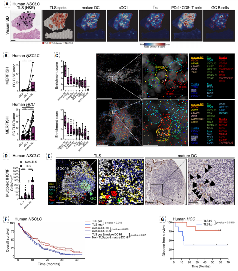
The study conducted panoramic analysis of multiple human solid tumors through multi-omics spatial mapping technology, clearly revealing the specific association between mature DCs and tumor TLSs, laying the clinical foundation for subsequent mechanism research. Spatial transcriptomics data showed that in pathological annotation TLS regions of tumors such as non-small cell lung cancer and hepatocellular carcinoma, characteristic genes of mature DCs, cDC1s, TFH cells, TPEX cells, and germinal center B cells were significantly enriched, while the tumor core regions lacked such immune enrichment characteristics. Macrophages and monocytes showed no obvious region-specific distribution, demonstrating that TLS is a specific immune aggregation microenvironment dominated by mature DCs. From the perspective of cell maturation characteristics, cDC1s within TLS expressed higher levels of maturation-related genes compared to cDC2s, suggesting that cDC1s are the core mature DC subset in TLS that loads tumor antigens and possesses immune activation functions.
Single-cell spatial interaction analysis further revealed that mature DCs in TLS can form proximal cell interactions with naive T/B cells, germinal center B cells, TFH cells, and TPEX cells, and are mainly localized in T cell regions or high-endothelial venule surroundings at the T-B cell boundary, replicating the immune cell distribution characteristics of secondary lymphoid organs. From the perspective of clinical prognosis, survival data from the POPLAR cohort confirmed that in non-small cell lung cancer patients, the population with high TLS density and high mature DCs characteristics had significantly prolonged overall survival; the BIONIKK renal cancer cohort and hepatocellular carcinoma neoadjuvant PD-1 treatment cohort also verified that patients with TLS enrichment accompanied by mature DCs infiltration showed better immunotherapy responses and longer disease-free survival, fully demonstrating that the infiltration level of mature DCs can serve as a core biomarker for tumor TLSs functional activity and patient prognosis.
3.2 Mature cDC1s are the Core Regulatory Cells for Murine Tumor TLSs Formation
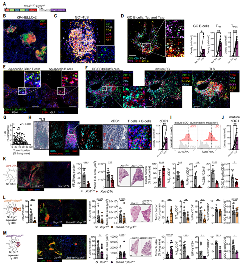
Using the KP-HELLO-2 mouse lung cancer model, the study precisely verified the decisive role of cDC1s in tumor TLSs development. The lung tumors of this mouse model can spontaneously form mature TLSs with intact structure, featuring clear T, B cell compartments, activated germinal centers, and enrichment of TFH cells, TPEX cells, and tumor-specific immune cells, showing high consistency with the structure and cellular composition of human tumor TLSs, demonstrating excellent model reliability. Experiments found that mature cDC1s in mouse tumor TLS highly expressed activation markers such as CCR7, MHC-II, CD86, can form tight cell interactions with T and B cells, and the area of TLS showed significant negative correlation with tumor burden, directly confirming the anti-tumor immune protective effect mediated by TLS.
Cell ablation experiments directly confirmed the core function of cDC1s. After specific depletion of cDC1s in Xcr1-DTA, Batf3-/- mice, the number and volume of TLSs in tumors decreased significantly, infiltration levels of TFH cells, IFN-γ+CD4+T cells, CD8+T cells, and germinal center B cells were significantly reduced, tumor burden increased markedly, and the remaining lymphoid aggregation structures completely lost the functional architecture of mature TLSs. Meanwhile, Wdfy4 gene deficiency leading to impaired cDC1s cross-presentation function resulted in blocked TLSs formation and impaired induction of tumor-specific CD8+T cells, demonstrating that the antigen cross-presentation capacity of cDC1s is a necessary condition for TLSs formation. Further mechanism exploration revealed that the IFN-γ receptor signaling of cDC1s is key to their maturation and activation; cDC1s-specific IFN-γ receptor knockout would directly inhibit DCs maturation and block TLSs development; while the loss of cell migration function mediated by CCR7 molecules would also disrupt the normal formation of TLSs, clarifying that IFN-γ-dependent maturation activation and CCR7-dependent migration localization are the two prerequisite core conditions for cDC1s to regulate TLSs formation.
3.3 In Situ Enrichment of cDC1s in Late Tumor Progression Maintains TLSs Functional Homeostasis
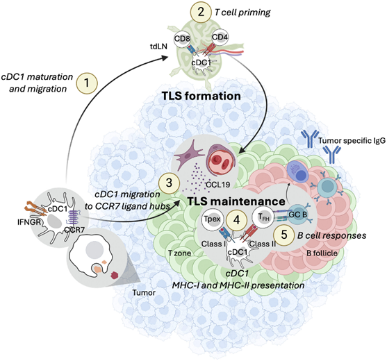
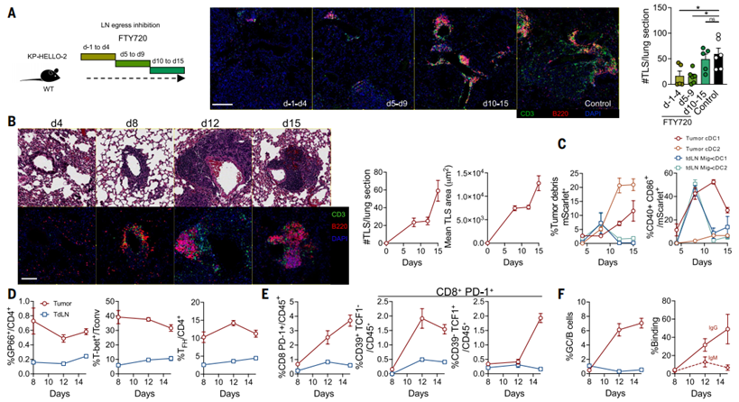
This study made a breakthrough in revealing the spatiotemporally differentiated regulatory mechanisms of TLSs formation and maintenance, overturning the previous understanding that TLSs fully depend on lymph node T cell output. Through temporal intervention experiments, the study found that in early tumor growth (within 9 days after inoculation), T cell activation output from tumor-draining lymph nodes was key to TLSs formation; at this time, blocking lymph node T cell egress would significantly inhibit TLSs generation; however, in late tumor progression, the homeostatic maintenance of TLSs completely detached from the continuous supply of lymph node T cells, showing local autonomous regulatory characteristics.
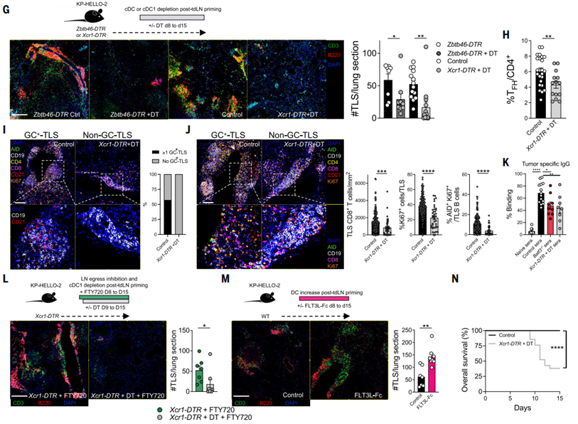
Dynamic lineage monitoring showed that in early tumor growth, mature cDC1s mainly migrated to tumor-draining lymph nodes to complete antigen presentation and T cell activation; while after 8 days of tumor growth, the migration trajectory of cDC1s underwent a significant shift, no longer accumulating in large quantities in draining lymph nodes, but continuously accumulating in the tumor in situ tissue. This transition was highly synchronized with the mature expansion of TLSs, differentiation of TPEX cells, germinal center formation, and tumor-specific IgG antibody production. To verify the function of in situ cDC1s, the study specifically ablated cDC1s at 8 days after tumor inoculation, and found that the already formed mature TLSs rapidly disintegrated, the TFH cell pool was substantially reduced, germinal center structures disappeared, tumor-specific IgM, IgG antibody secretion significantly decreased, CD8+T cell immune responses were impaired, directly proving that tumor in situ cDC1s are the core hub for late TLSs structure maintenance and functional activation. Conversely, FLT3L-Fc drug intervention can effectively expand in vivo cDC1s numbers, significantly increasing TLSs density in tumors, providing direct therapeutic evidence for targeting DCs to enhance tumor local immunity and activate TLSs anti-tumor function. Meanwhile, cDC1s ablation also significantly shortened the survival of tumor-bearing mice, confirming the anti-tumor protective effect of the cDC1s-TLS axis.
3.4 CCR7-CCL19 Axis-Mediated In Situ Localization of cDC1s Supports TLSs Maintenance
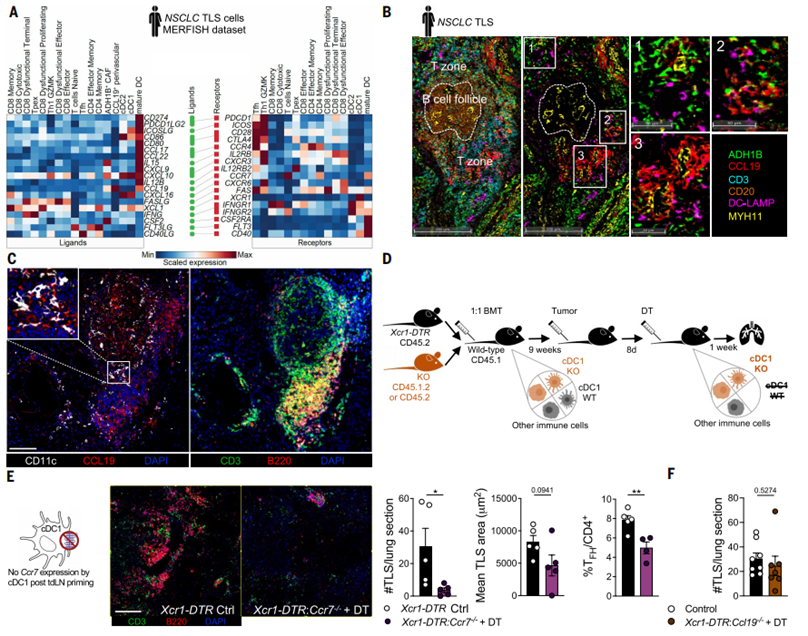
The study further elucidated the microenvironmental molecular mechanisms of tumor in situ cDC1s precise localization and TLSs maintenance. Through single-cell ligand--receptor interaction mapping and spatial imaging validation of human non-small cell lung cancer, it was found that cancer-associated fibroblasts and perivascular stromal cells in the tumor microenvironment can specifically secrete CCL19, forming stromal microenvironment hotspots enriched with CCR7 ligands, while mature cDC1s highly express CCR7 receptors, enabling precise chemotactic aggregation at these stromal hotspots, ultimately localizing in the core regions of TLSs. This specific spatial localization pattern was also verified in mouse tumor models, where mouse TLS regions were highly enriched with CCL19 signals, and cDC1s specifically aggregated in this region and maintained TLS structural stability.
Temporal gene knockout experiments precisely verified the function of the CCR7-CCL19 axis. After specific knockout of the CCR7 gene in cDC1s in late tumors, cDC1s were unable to migrate and enrich toward tumor stromal CCL19 hotspots, leading to reduced TLSs numbers, atrophied volumes, significantly decreased TFH cell infiltration, and complete disruption of the intact immune architecture of TLSs. Meanwhile, the study ruled out the role of cDC1s themselves secreting CCL19, confirming that stromal cell-derived CCL19 is the core signal regulating cDC1s localization and maintaining TLSs homeostasis, clarifying that "stromal CCL19-cDC1s CCR7 chemotactic localization" is the key spatial regulatory pathway for local TLSs maintenance in late tumors.
3.5 cDC1s Dual Antigen Presentation Pathways Synergistically Regulate TLSs Functional Homeostasis
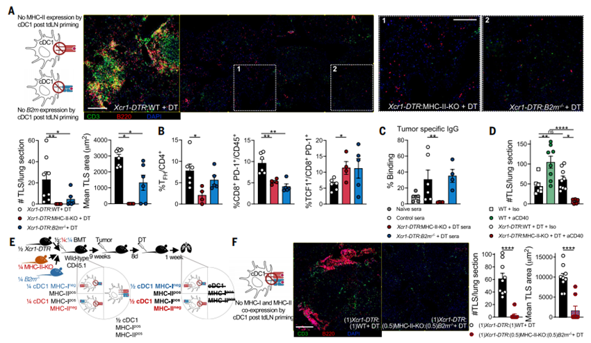
This study ultimately elucidated the core molecular mechanism of cDC1s maintaining TLSs structure and function, namely through the synergistic regulation of multiple tumor local immune responses via MHC-I and MHC-II dual antigen presentation pathways. Through conditional gene knockout, the study separately blocked the MHC-II and MHC-I antigen presentation functions of cDC1s, and found that the two pathways played differentiated yet complementary regulatory roles. After specific knockout of the MHC-II gene in cDC1s, TLSs structures were severely damaged, T and B cells were diffusely distributed, the TFH cell pool was substantially depleted, tumor-specific IgG antibody production was blocked, CD8+T cell activation was impaired, and TPEX cells abnormally accumulated, demonstrating that MHC-II-mediated CD4+T cell antigen presentation is the core basis for maintaining TFH cell homeostasis, germinal center formation, humoral immune response, and intact TLSs structure.
After specific knockout of the MHC-I gene in cDC1s and blocking of antigen cross-presentation, the structural damage to TLSs was relatively moderate, TFH cell numbers and tumor antibody secretion showed no significant changes, but the activation and expansion of CD8+T cells were significantly inhibited, and TPEX cell differentiation was imbalanced, confirming that MHC-I cross-presentation is mainly responsible for regulating the anti-tumor immune function of CD8+T cells and is key to TLSs-mediated cellular immune responses. Additionally, the study found that CD40 signaling is an important auxiliary pathway for cDC1s to exert antigen presentation functions, and cDC1s-specific CD40 deficiency would significantly weaken TLSs homeostasis. More critically, the pro-TLSs expansion effect of CD40 agonists completely depends on the MHC-II antigen presentation function of cDC1s. When the MHC-II pathway was blocked, drug intervention was completely ineffective, ultimately confirming that cDC1s synergistically maintain TLSs structural integrity and functional activation through simultaneously activating MHC-II-mediated CD4+T cell responses and MHC-I-mediated CD8+T cell responses, building the core framework of tumor local adaptive immune responses.
IV. Research Summary
This study systematically and comprehensively elucidated the spatiotemporal molecular mechanisms of cDC1s regulating tumor tertiary lymphoid structure formation, maintenance, and functional activation, breaking the previous one-sided understanding of TLSs regulatory mechanisms and establishing a dynamic regulatory model of tumor TLSs centered on mature cDC1s. The overall study confirmed that the development and homeostatic maintenance of tumor TLSs exhibit clear temporal differentiation: early tumors depend on IFN-γ signal-driven cDC1s maturation, CCR7-mediated lymph node migration, and draining lymph node T cell output to initiate the initial formation of TLSs; in late tumor progression, it completely switches to a tumor in situ autonomous regulatory mode, relying on the stromal cell CCL19-CCR7 signaling axis to guide cDC1s specific enrichment in the tumor locality, through cDC1s simultaneously activating MHC-II and MHC-I dual antigen presentation pathways, respectively maintaining CD4+ TFH cell homeostasis, humoral immune responses, and CD8+T cell anti-tumor cellular immunity, supporting TLSs structural integrity and long-term functional persistence. Meanwhile, the study combined multiple human tumor samples and clinical cohorts to verify the high correlation of mature cDC1s, TLSs with favorable prognosis in tumor patients and immune checkpoint therapy response, and through intervention experiments with FLT3L-Fc, CD40 agonists, and other interventions, confirmed that targeted activation and expansion of cDC1s can effectively enhance TLSs number and function, providing novel targets and intervention strategies for tumor immunotherapy. Overall, this study established that cDC1s are the core organizer and functional regulatory hub of tumor TLSs, perfected the molecular mechanisms of tumor local adaptive immune activation, and provided solid theoretical and experimental support for subsequent optimization of TLSs-targeted immunotherapy and improvement of solid tumor immunotherapy efficacy.
EnkiLife not only provides customers with a complete set of TSA multiplex labeling kits, but also offers various TSA specialty technical services, including IF fluorescence staining, fluorescence panoramic scanning, ultra-multiplex staining, and pathological analysis (5 markers and below).
Product | Catalog Number |
|---|---|
7-Color Multiple Fluorescent Staining Kit (mIHC) | |
6-Color Multiple Fluorescent Staining Kit (mIHC) | |
5-Color Multiple Fluorescent Staining Kit (mIHC) | |
4-Color Multiple Fluorescent Staining Kit (mIHC) | |
3-Color Multiple Fluorescent Staining Kit (mIHC) |
For details, please check TSA mIHC Kit