Adipogenic Induction
Obesity is characterized by an increase in the mass or dysfunction of adipose tissue resulting from an imbalance between energy intake and expenditure; it is closely associated with metabolic disorders such as diabetes, cardiovascular disease, non-alcoholic fatty liver disease, and certain cancers. The expansion of adipose depots—particularly white adipose tissue—is characterized by either an increase in adipocyte volume (hypertrophy) or the formation of new adipocytes from precursor cells (hyperplasia). Consequently, the induction of cellular adipogenesis plays a pivotal role in mechanistic research and drug development related to obesity-associated diseases.
Adipogenic differentiation is the process by which mesenchymal stem cells (MSCs) or pre-adipocytes are induced to differentiate into mature adipocytes under specific conditions; the extent of this differentiation can be assessed using Oil Red O staining. The 3T3-L1 cell line is the most widely used model for studying adipogenic differentiation.
Tips:
Adipose tissue can be broadly classified into white adipose tissue and brown adipose tissue.
Mature white adipocytes contain numerous large, unilocular lipid droplets; these cells are polygonal in shape, feature a peripherally located nucleus, and contain relatively few mitochondria. Key marker genes include Agt, Retn (Resistin), Slc2a4 (GLUT4), Cfd (Adipsin), Adipoq (Adiponectin), and Fabp4 (aP2).
Brown adipocytes contain multiple small, multilocular lipid droplets and are mitochondria-rich cells specialized for thermogenesis. Key marker genes include DIO2, PPARα, PRDM16, and UCP1.
Cell Selection:
3T3-L1: Exhibits excellent adipogenic potential and reproducibility; however, as a murine-derived cell line, it presents certain differences compared to human-derived cells.
hMSCs: Human bone marrow-derived MSCs, offering high clinical relevance; however, their differentiation efficiency is relatively low (approximately 30%).
hADSCs: Human adipose-derived mesenchymal stem cells, characterized by high differentiation efficiency; however, they exhibit significant variability depending on the individual donor.
Adipogenic Differentiation Protocol (Using Product RC0044 to Induce 3T3-L1 Cells as an Example)
1. Harvest cells in the logarithmic growth phase and seed them into culture vessels at a density of 2.0 × 10⁴ cells/cm². Incubate the cells at 37°C in a 5% CO₂ atmosphere until they reach 90–100% confluence.
2. Discard the supernatant and add the Adipogenic Induction Medium (Induction Solution). Incubate at 37°C in a 5% CO₂ atmosphere for approximately 3 days. Then, replace the medium with the Adipogenic Induction Medium (Maintenance Solution) and incubate for 1 day. Subsequently, switch back to the Adipogenic Induction Medium (Induction Solution) and continue incubating for another 3 days. Continue this cycle of medium exchange for a total induction period of 14–21 days, carefully observing changes in cell morphology throughout the process. Determine the appropriate time to terminate the induction based on the quantity and size of the lipid droplets formed within the cells, and proceed with staining for identification.
3. Aspirate the culture medium and wash the cells once with an appropriate volume of 1× PBS. Discard the wash solution, then add a sufficient volume of 4% Paraformaldehyde (PFA) solution to cover the bottom of the culture vessel. Fix the cells at room temperature for 30–60 minutes. Discard the fixing solution and wash the cells twice with 1× PBS.
4. Prepare the Oil Red O working solution by mixing the Oil Red O stock solution with either physiological saline or 1× PBS (at a ratio of 3:2). Filter the solution immediately after preparation; the working solution should be prepared fresh just before use. Add an appropriate volume of the Oil Red O working solution to the washed induction wells and allow the cells to stain undisturbed for 30 minutes. Aspirate the Oil Red O working solution, wash the cells twice with 1× PBS, and add a sufficient volume of 1× PBS to prevent the cells from drying out.
5. Observe the results of the adipogenic staining under a microscope to capture images and evaluate the success of the induction. If the induction was successful, the lipid droplets will appear red or orange-red after binding with the Oil Red O dye.
Images from Adipogenic Induction using EnkiLife Adipogenic Induction Medium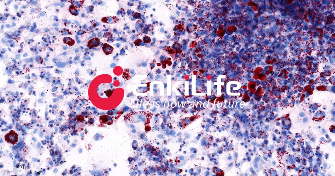
Figure 1. Oil Red O staining results following adipogenic induction_20.0x-1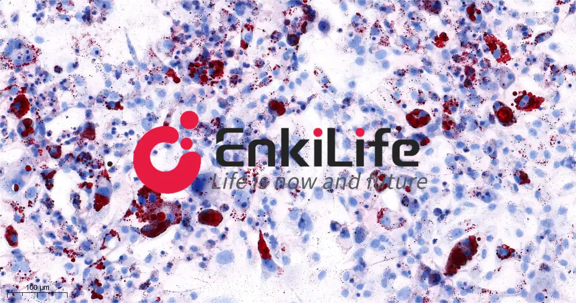
Figure 2. Oil Red O staining results following adipogenic induction_20.0x-2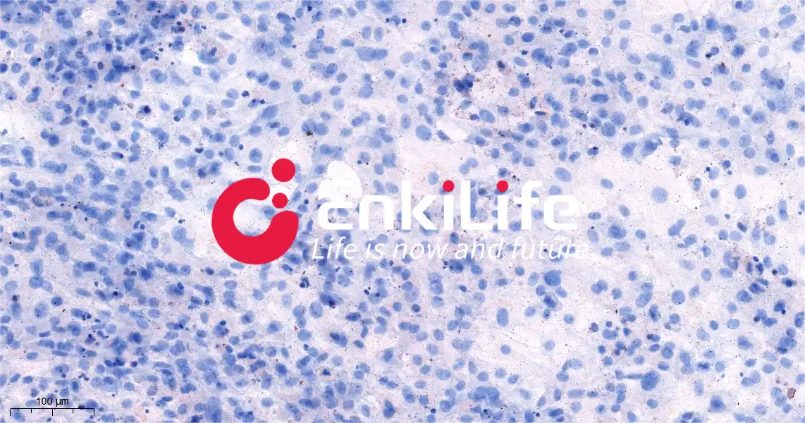
Figure 3. Oil Red O staining results without adipogenic induction_20.0x-1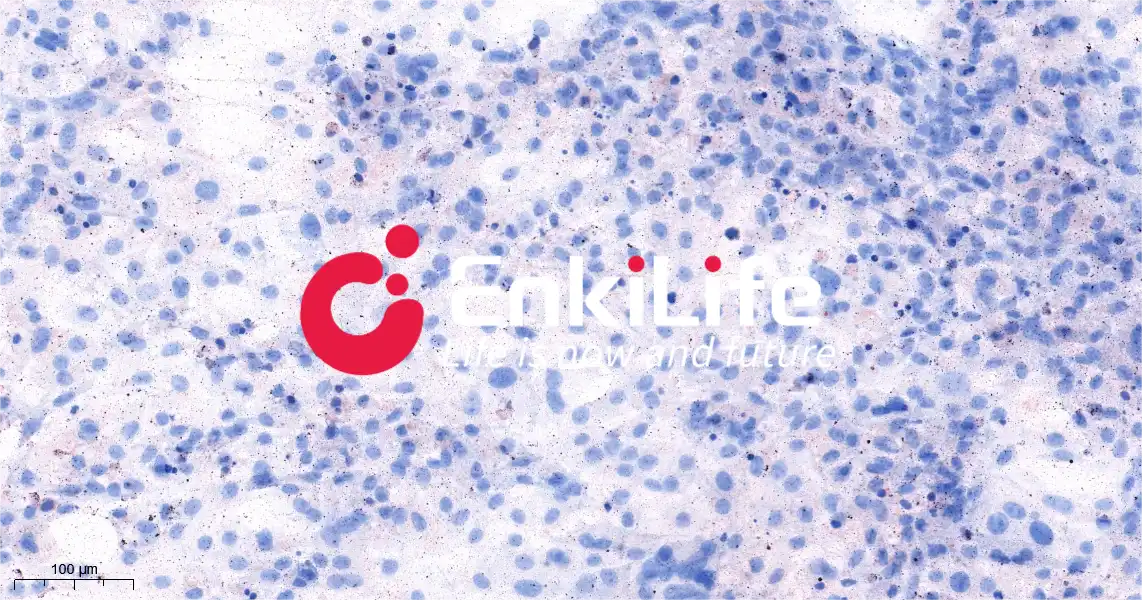
Figure 4. Oil Red O staining results without adipogenic induction_20.0x-2
Common Issues and Troubleshooting
1. Insufficient Lipid Droplet Formation
Insufficient cell density: Failure to reach contact inhibition leads to induction failure. When performing adipogenic induction, ensure that cells have reached a density of 90–100% before initiating the induction process.
Inactive induction reagents: Reagent degradation leads to induction failure. Insulin must be stored away from light, and repeated freeze-thaw cycles should be avoided; Dexamethasone must also be stored away from light.
Over-fixation with 4% Paraformaldehyde (PFA): Excessive fixation time can damage lipid droplet structures. The standard fixation time is typically 30–60 minutes; it is recommended to optimize the fixation time based on observed results.
Crystallization of Oil Red O staining solution: Crystal formation in the stain leads to staining failure. The Oil Red O solution must be filtered after preparation before use. Additionally, saturated staining solutions do not stain effectively; therefore, the Oil Red O solution must be diluted strictly according to the instructions provided in the product manual.
Insufficient induction duration: An inadequate induction period results in a lack of lipid droplets. For certain cell types—such as umbilical cord mesenchymal stem cells—the induction period may exceed 30 days, and lipid droplet formation can be difficult to observe. The induction duration should be optimized based on the specific cell type being used.
2. Excessive Background Staining
Insufficient washing: Inadequate washing leads to a dark background. Optimize the number of washes and the washing duration; additionally, after the staining process is complete, a brief wash with 60% Isopropanol for 10 seconds may be applied to reduce background staining.
Excessive staining duration: Prolonged staining time exacerbates background staining. Oil Red O staining time should generally be limited to within 30 minutes; a pilot experiment can be performed to determine the optimal staining duration.
3. Significant Cell Death
Poor cell health: Failure to change the culture medium in a timely manner during the induction period can lead to cell death. Assess the condition of the cells prior to plating, and use Trypan Blue staining to determine cell viability.
Rough handling during medium exchange: Aggressive manipulation while changing the medium can cause physical damage to the cells, resulting in cell death. Handle cells gently, and when changing the medium, slowly dispense the fresh medium along the inner wall of the culture well.
🔎3T3-L1 Adipogenic Differentiation Medium Kit:https://enkilife.com/p/3T3-L1-Adipogenic-Differentiation-Medium-Kit-RC0044
📍 Get in touch: order@enkilife.com
 | Flora Flora is a technical support expert at EnkiLife, familiar with immunology and neuroscience, dedicated to providing customers with high-quality product combinations and technical support to help achieve research in neurodegenerative diseases and other neuroscience areas. |