Literature Sharing: Combined Multiplex Immunofluorescence and Traditional Histology Imaging for Cancer Biomarker Discovery
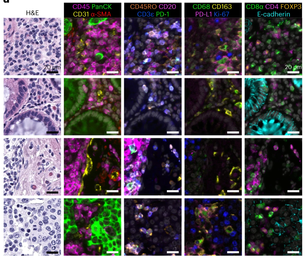
Precise analysis of the tumor microenvironment is the core foundation for cancer diagnosis, prognosis assessment, and treatment decision-making. However, traditional tissue imaging techniques have obvious limitations: single immunofluorescence (IF) imaging can detect multiple molecular markers but lacks histological context, making it difficult to accurately localize cell types and distributions. Hematoxylin-Eosin (H&E) staining, as the gold standard for pathological diagnosis, clearly presents tissue morphology but cannot provide molecular-level immune characteristic information. Existing multiplex imaging techniques either have complex processes and are time-consuming, or have issues such as fluorescence crosstalk and insufficient reagent stability, making it difficult to achieve simultaneous precise detection of molecular markers + tissue morphology.