Fluorescent Dye or Marker Characteristics
We offer a variety of small-molecule fluorescent dyes (typically referring to fluorescent dyes with a molecular weight of around 1000D), represented by cyanine series and Fluor series dyes. These are suitable for most detection channels/filters in various testing instruments, and many small-molecule fluorescent dyes can be used for multiplex detection. This series of fluorescent dyes covers near-ultraviolet, visible light, and near-infrared spectra, and is compatible with common excitation light sources and instruments. Below, we provide detailed introductions to some dyes, which can serve as a reference for users selecting fluorescent dye antibody protein labeling kits, fluorescent secondary antibodies, flow cytometry antibodies, etc.
Fluorescent Dye Characteristics Comparison Table
| Emission Light Name | Dye Name | Common Applications | Ex max/Em max | Equivalent/Approximate Spectrum Dyes |
|---|---|---|---|---|
| Blue | Fluor350 | IF Imaging | 346nm/445nm | AMCA, Coumarin |
| Blue-Purple | Fluor405 | FC,IF Imaging | 400nm/424nm | |
| Green | Cyanine2 | IF Imaging | 490nm/510nm | |
| FITC | FC,IF Imaging | 495nm/520nm | ||
| Fluorescein-X | FC | 495nm/519nm | ||
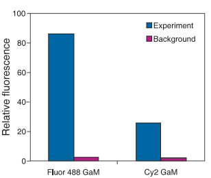
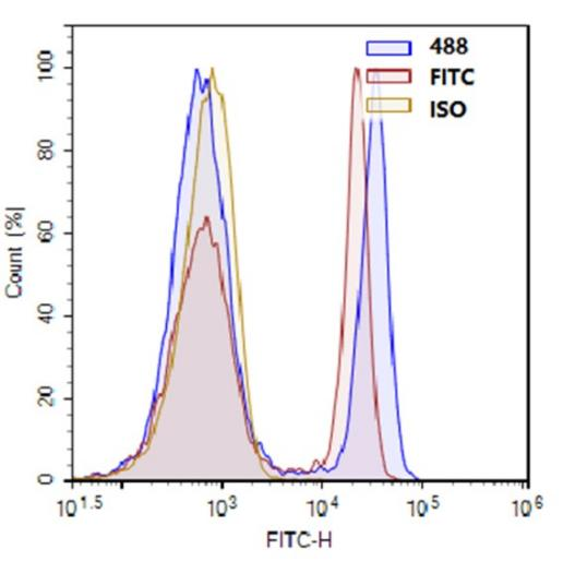
| Fluor488 | FC,IF Imaging | 490nm/513nm | Cy2, Fluorescein-X,FITC (Fluorescein) | |
| Yellow-Green | Fluor430 | IF Imaging | 430nm/545nm | |
| Fluor532 | IF Imaging | 530nm/555nm | ||
| Yellow | Cyanine3 | IF Imaging | 548nm/563nm | |
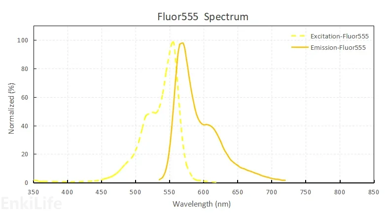
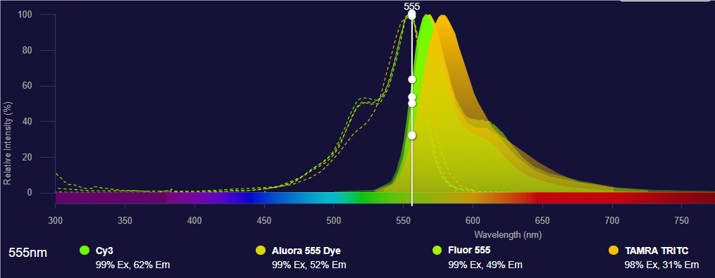
| Fluor555 | IF Imaging | 555nm/572nm | Cy3, TRITC (Rhodamine) | |
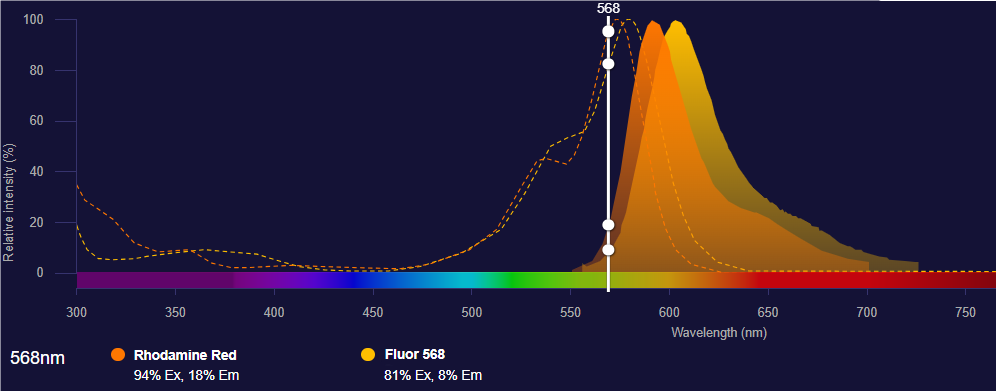
| Orange | Fluor568 | IF Imaging,FC | 578nm/602nm | Rhodamine Red |
| Fluor590 | IF Imaging | 590nm/620nm | ||
| Fluor594 | IF Imaging,FRET | 590nm/617nm | Texas Red | |
| Texas Red | Fluor647 | FC,IF Imaging | 651nm/668nm | Cy5,APC |
| Cyanine5 | IF Imaging | 646nm/662nm | ||
| Cyanine5.5 | IF Imaging,FRET | 675nm/694nm | ||
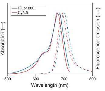
| Near Red | Fluor680 | IF Imaging | 680nm/702nm | Cy5.5, IR680 |
| Fluor700 | FC | 702nm/723nm | ||
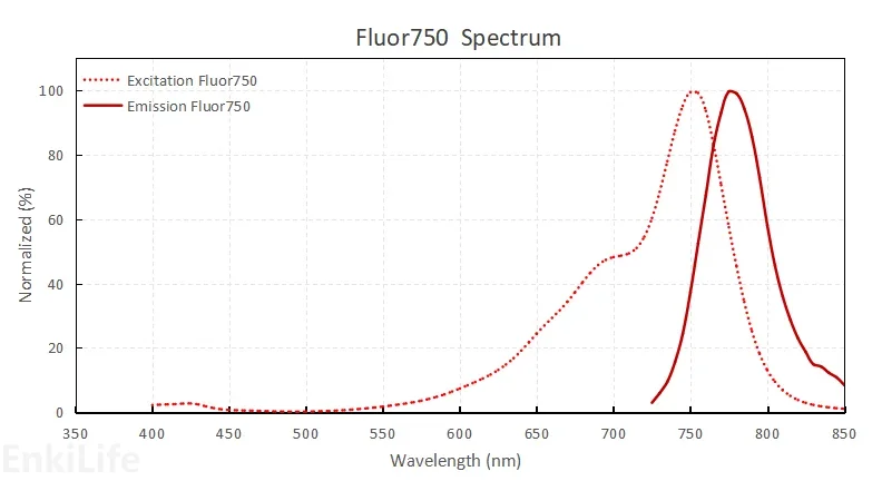
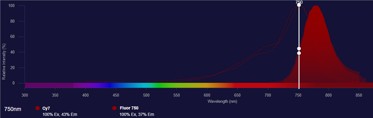
| Fluor750 | IF Imaging | 747nm/770nm | Cy7 | |
| Cyanine7 | IF Imaging,FRET | 750nm/773nm | ||
| Fluor770 | WB Fluorescence | 770nm/790nm |
Fluorescent Dye or Marker Characteristics Introduction
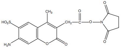
Fluor350
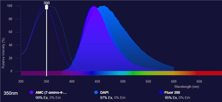
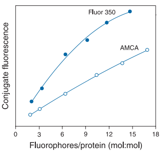
Fluor350 is an excellent blue fluorescent dye, a modified dye based on the coumarin dye 7-AMCA skeleton. It can serve as an excellent alternative to AMCA, commonly used in fluorescence imaging and flow cytometry analysis, with good water solubility and pH insensitivity. The excitation and emission spectra of Fluor350 are close to those of DAPI, and it can be directly imaged using DAPI filter sets.

Application References:
- Rothschild, K. J.; Gite, S.; Olejnik, J. Methods for the detection, analysis and isolation of nascent proteins. U.S. Pat. Appl. Publ. US 20110250609,2011.
- Zauner, G.; Lonardi, E.; Bubacco, L.; Aartsma,T. J.; Canters, G. W.; Tepper, A. W. J. W.Tryptophan-to-dye fluorescence energy transferapplied to oxygen sensing by using type-3 copper proteins. Chem.- Eur. J. 2007, 13, 7085--7090.
- Gee, K. R.; Hart, C. CR.; Haugland, R.; Patton,W. F.; Whitney, S. Site-specific labeling of affinitytags in fusion proteins. U.S. Pat. Appl. Publ. US.20060141554, 2006.
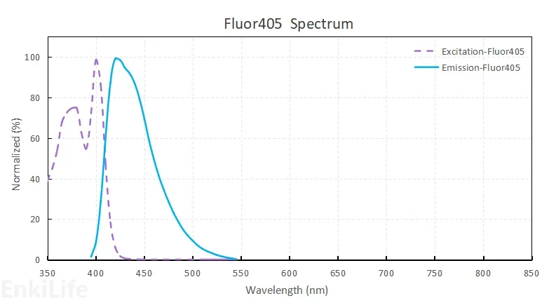
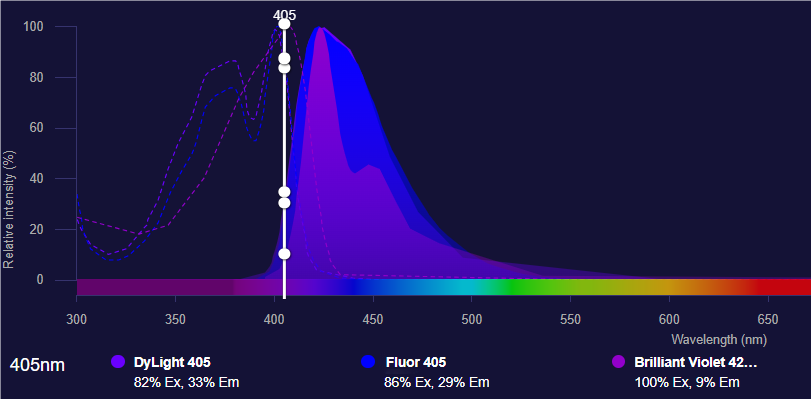
Fluor405
The excitation/emission maximum of Fluor405 blue fluorescent dye is 400/424nm, which almost perfectly matches the spectral lines of the 405nm violet laser widely used in fluorescence microscopy and flow cytometry. The excitation and emission spectra of Fluor405 highly overlap with those of BV421 dye. Fluor405 is a derivative of Cascade Blue dye, but with optimized fluorescence properties after structural modification. The spectral overlap between Fluor405 dye and green fluorophores is minimal, making it an ideal choice for multicolor applications.
Application References:
- Jones SA, Shim SH, He J, Zhuang .Fast, three-dimensional super-resolution imaging of live cells.Nat Methods. 2011 Jun;8(6):499-508.
- Camila van Zanten , Dzmitry Melnikau , Alan G Ryder.Effects of Viscosity and Refractive Index on the Emission and Diffusion Properties of Alexa Fluor 405 Using Fluorescence Correlation and Lifetime Spectroscopies.J Fluoresc. 2021 May;31(3):835-845.
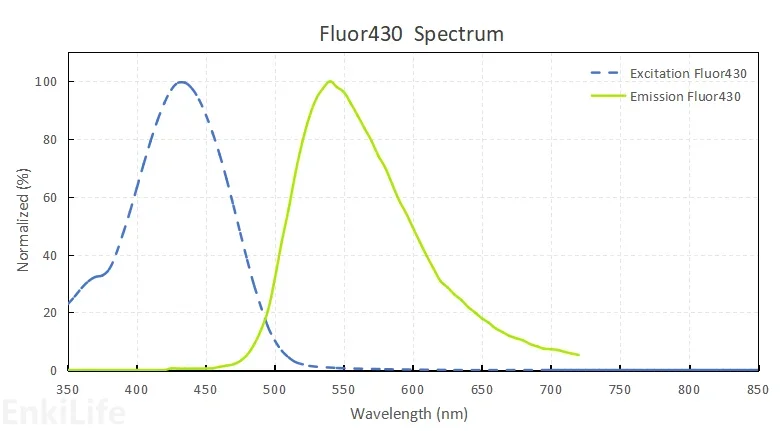
Fluor430
Among active dyes that absorb light in the wavelength range of 400-450nm, significant fluorescence is usually not produced above 500nm. The Fluor430 dye fills this gap in the wavelength range. When excited near its absorption peak at 430nm, this dye exhibits a large Stokes shift and intense yellow-green fluorescence.
Application References:
- Tsilivakos, V.; Gritzapis, A. Method of intracellular infectious agent detection in sperm cells. PCT Int.Appl. WO 2013144662, 2013.
- Arcangeli, A.; Becchetti, A.; Pillozzi, S.; Masselli,M.; De Lorenzo, E. Method and kit for the prevention and/or the monitoring of chemoresistance of leukemia forms. PCT Int. Appl. WO 2011058509,2011.
- Lewis B, Rathman S, McMahon RJ.Detection and quantification of biotinylated proteins using the Storm 840 Optical Scanner.J Nutr Biochem.2003 Apr;14(4):196-202.
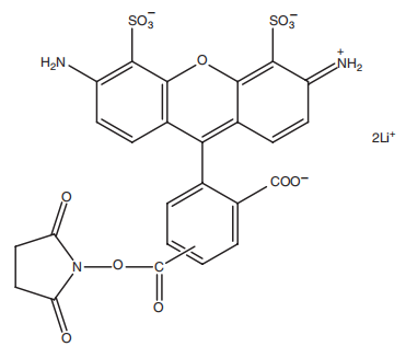
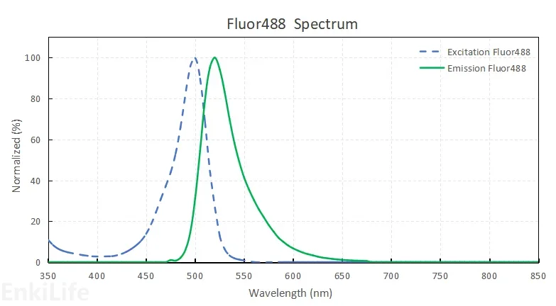
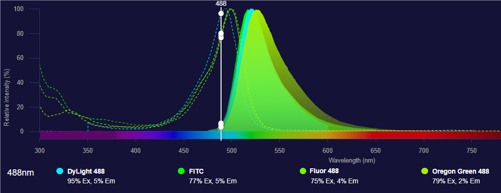
Fluor488
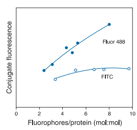
Fluor488 is sulfonated rhodamine green. By introducing two sulfonate ions, Fluor488 has significantly enhanced water solubility, pH stability, and photostability. Unlike fluorescein dyes, the fluorescence of Fluor488 remains unchanged over a wide pH range (4-10). Due to its multiple negative charges, Fluor488 is less prone to intermolecular aggregation, and when multiple dye molecules label the same protein, it is less prone to fluorescence self-quenching or causing protein aggregation. Fluor488 is an excellent substitute for fluorescein (FITC or FAM) and CY2, performing extremely well in single-molecule detection of bioconjugates, fluorescence correlation spectroscopy, and fluorescence polarization measurements. It is also widely used in fluorescence microscopy and flow cytometry. Fluor488 is currently one of the best water-soluble small-molecule green fluorescent dyes.
Application References:
- Karver, M. R.; Weissleder, R.; Hilderbrand, S. A.Bioorthogonal reaction pairs enable simultaneous,selective, multi-target imaging. Angew. Chem., Int.Ed. 2012, 51, 920--922.
- Lundberg, E.; Sundberg, M.; Graeslund, T.; Uhlen,M.; Svahn, H. A. A novel method for reproducible fluorescent labeling of small amounts of antibodies on solid phase. J. Immunol. Methods 2007, 322,40--49.
- Comparison between photostability of Alexa Fluor 448 and Alexa Fluor 647 with conventional dyes FITC and APC by flow cytometry.Int J Lab Hematol. 2018 Jun;40(3):e52-e54.
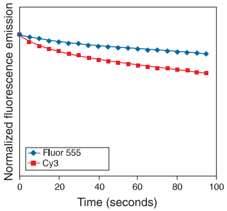
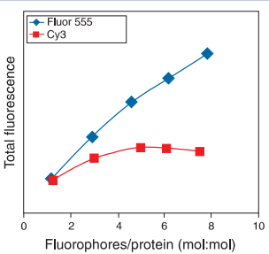
Fluor555
Fluor555 is an orange-orange-red fluorescent dye, an analog of Cyanine3, commonly used to replace Cyanine3 and TAMRA dyes. Fluor555 dye has high polarity, strong water solubility, excellent fluorescence brightness and fluorescence stability, making it a good dye for protein/antibody labeling. From a chemical structure perspective, the parent nucleus of Fluor555 is cyanine Cyanine3, but the parent nucleus has up to 4 sulfonic acid groups. The tetrasulfonic acid structure makes it extremely hydrophilic, and when the dye is labeled on the protein surface, it does not change the protein surface polarity, reducing the risk of protein hydrophobic aggregation and precipitation, and greatly reducing the probability of fluorescence quenching due to local proximity of the dye. Therefore, proteins labeled with Fluor555 often emit stronger fluorescence. However, because most flow cytometers currently on the market are traditional flow cytometers (not spectral flow cytometers), the laser compatibility is not optimal, so Fluor555 dye is more used in the field of fluorescence imaging.
Application References:
- Arcangeli, A.; Becchetti, A.; Pillozzi, S.; Masselli,M.; De Lorenzo, E. Method and kit for the prevention and/or the monitoring of chemo resistance of leukemia forms. PCT Int. Appl. WO 2011058509,2011.
- Lundberg, E.; Sundberg, M.; Graeslund, T.; Uhlen,M.; Svahn, H. A. A novel method for reproducible fluorescent labeling of small amounts of antibodies on solid phase. J. Immunol. Methods 2007, 322,40--49.
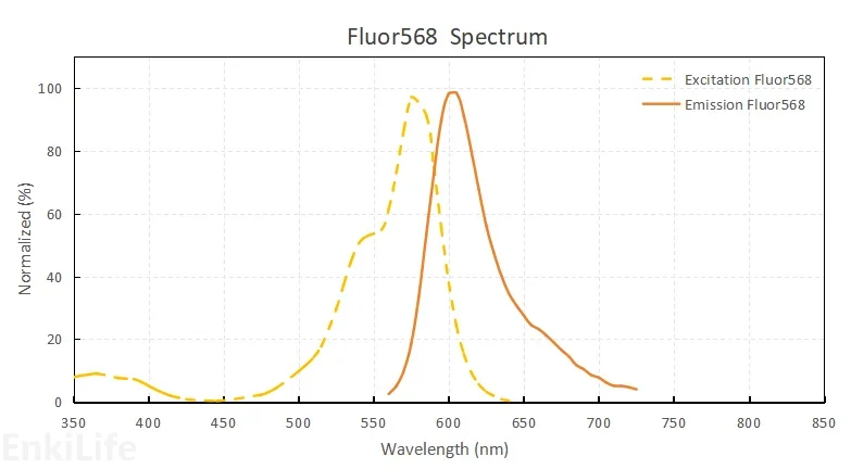
Fluor568
Fluor568 fluorescent dye perfectly matches 561nm lasers. The maximum excitation/emission of this red-orange fluorescent dye is approximately 578/602nm, which can achieve optimal excitation effect in the 561nm diode laser used in many confocal laser scanning microscopes. Fluor568 has a parent nucleus of rhodamine compounds, and two sulfonate ions are introduced to optimize the water solubility and optical properties of the dye. When excited, it emits bright orange-red fluorescence, which is very sensitive and stable, and can be used to detect low-abundance targets. It is an excellent water-soluble small-molecule fluorescent labeling dye.
Application References:
- Jafar Mahmoudian, Reza Hadavi, Mahmood Jeddi-Tehrani.Comparison of the Photobleaching and Photostability Traits of Alexa Fluor 568- and Fluorescein Isothiocyanate-conjugated Antibody.Cell J. 2011 Fall;13(3):169-72.
- Seyed Nasser Ostad, Sepideh Babaei , Ali Ahmad Bayat. Photobleaching Comparison of R-Phycoerythrin-Streptavidin and Streptavidin-Alexa Fluor 568 in a Breast Cancer Cell Line. Monoclon Antib Immunodiagn Immunother. 2019 Feb;38(1):25-29.
- Tamás Gajdos , Béla Hopp.Hot-Band Anti-Stokes Fluorescence Properties of Alexa Fluor 568.J Fluoresc.2020 May;30(3):437-443.
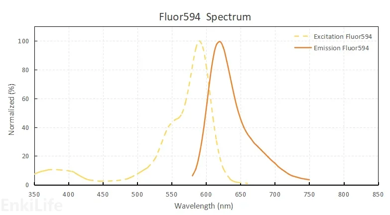
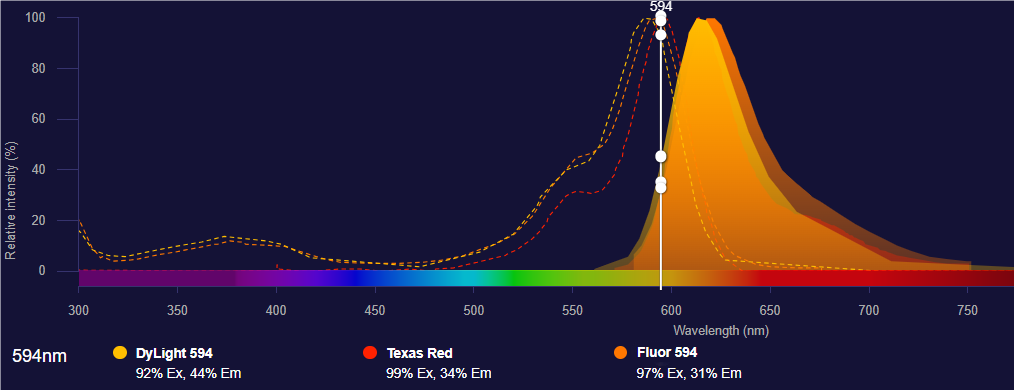
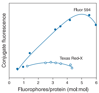
Fluor594
Fluor594 is a rhodamine compound methylated on the basis of Fluor568 structure. Two sulfonate ions also play a role in optimizing the properties of the dye. When excited, it emits bright red fluorescence, which is very sensitive and stable, and can be used to detect low-abundance targets. Fluor594 is an excellent water-soluble small-molecule fluorescent labeling dye, suitable for labeling various target molecules, and widely used in various biological experiments. Conjugates prepared with Fluor594 dye emit fluorescence in the red region of the spectrum, so they are particularly suitable for multiplex labeling experiments combined with green fluorescent probes. The spectral characteristics of Fluor594 are similar to those of Cyanine3.5 and Texas Red, but the fluorescence intensity is stronger, making it an excellent substitute for Texas Red.
Application References:
- Arcangeli, A.; Becchetti, A.; Pillozzi, S.; Masselli,M.; De Lorenzo, E. Method and kit for the prevention and/or the monitoring of chemo resistance of leukemia forms. PCT Int. Appl. WO 2011058509,2011.
- Green, D. P. L.; Rawle, C. B. Analysis system and method. PCT Int. Appl. WO 2009082242, 2009.
- N Panchuk-Voloshina , R P Haugland, J Bishop-Stewart. Alexa dyes, a series of new fluorescent dyes that yield exceptionally bright, photostable conjugates. J Histochem Cytochem. 1999 Sep;47(9):1179-88.
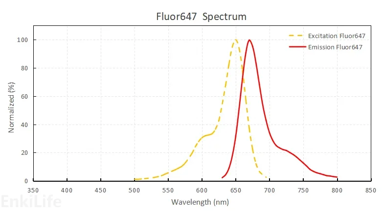
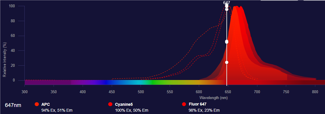
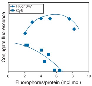
Fluor647
The spectrum of Fluor647 fluorescent dye almost completely matches that of Cyanine5, so the compatibility with optical filters designed for Cyanine5 can also be optimal. However, the total fluorescence intensity of Fluor647 antibody conjugates is usually significantly higher than that of Cyanine5 conjugates. Moreover, when Fluor647 is conjugated with most proteins, oligonucleotides, and nucleic acids, its absorbance or fluorescence spectrum shows almost no change, so under the same degree of substitution, the total fluorescence intensity is higher, making it an excellent substitute for Cyanine5.
Application References:
- Liming Ou, Qingyu Lv, Canjun Wu, Huaijie Hao. Development of a lateral flow immunochromatographic assay for rapid detection of Mycoplasma pneumoniae-specific IgM in human serum specimens.J Microbiol Methods. 2016 May:124:35-40.
- N Panchuk-Voloshina , R P Haugland, J Bishop-Stewart. Alexa dyes, a series of new fluorescent dyes that yield exceptionally bright, photostable conjugates. J Histochem Cytochem. 1999 Sep;47(9):1179-88.
- Ryo Tsumura , Ryuta Sato , Fumiaki Furuya.Feasibility study of the Fab fragment of a monoclonal antibody against tissue factor as a diagnostic tool.Int J Oncol. 2015 Dec;47(6):2107-14.
- Comparison between photostability of Alexa Fluor 448 and Alexa Fluor 647 with conventional dyes FITC and APC by flow cytometry.Int J Lab Hematol. 2018 Jun;40(3):e52-e54.
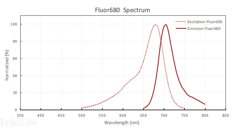
Fluor680
The peak excitation wavelength of Fluor680 dye is 680nm, and the maximum emission wavelength is 702nm. Its spectral characteristics are similar to Cyanine5.5 dye. The fluorescence of Fluor680 dye is clearly separated from the emission signals of other commonly used red fluorescent dyes (such as tetramethylrhodamine, R-phycoerythrin, Fluor594, and Fluor647), so it is very suitable for multicolor labeling.
Application References:
- Ogawa, M.; Regino, C. A.; Choyke, P. L.; Kobayashi,H. In vivo target-specific activatable near-infrared optical labeling of humanized monoclonal antibodies.Mol. Cancer Ther. 2009, 8, 232--239.
- N Panchuk-Voloshina , R P Haugland, J Bishop-Stewart. Alexa dyes, a series of new fluorescent dyes that yield exceptionally bright, photostable conjugates. J Histochem Cytochem. 1999 Sep;47(9):1179-88.
Fluor750
Fluor750 is spectrally very similar to Cyanine7 dye, and is an excellent substitute for Cyanine7. The maximum fluorescence emission wavelength is 770nm, which is far from the maximum emission wavelength of commonly used far-red fluorescent substances (Fluor647 or APC), making it convenient for multicolor analysis.
Application References:
- Paris-Robidas, S.; Emond, V.; Tremblay, C.; Soulet,D.; Calon, F. In vivo labeling of brain capillary endothelial cells after intravenous injection ofmonoclonal antibodies targeting the transferrin receptor. Mol. Pharmacol. 2011, 80, 32--39.
- Paudyal, P.; Paudyal, B.; Iida, Y.; Oriuchi, N.;Hanaoka, H.; Tominaga, H.; Ishikita, T.; Yoshioka,H.; Higuchi, T.; Endo, K. Dual functional molecular imaging probe targeting CD20 with PET and optical imaging. Oncol. Rep. 2009, 22, 115--119.
- Moinuddin Hassan 1, Victor Chernomordik, Rafal Zielinski.In vivo method to monitor changes in HER2 expression using near-infrared fluorescence imaging.Mol Imaging.2012 Jun;11(3):177-86.
 | Hansun Hansun is EnkiLife's protein labeling expert, proficient in immunology, various labeling techniques, and flow cytometry detection. We are committed to the precise labeling of proteins to empower scientific and technological breakthroughs. Every protein deserves to be "seen". |