Multiplex Immunofluorescence (mIHC) Technology
Traditional pathology has long been the cornerstone of disease diagnosis, relying on qualitative or semi-quantitative visual examination of tissue sections to detect pathological changes. While single immunohistochemistry (IHC) can effectively detect specific biomarkers, it is often limited to a single biomarker, which restricts its ability to capture the complexity of the tissue environment. The introduction of multiplex imaging technologies, such as multiplex IHC and multiplex immunofluorescence (mIHC), represents a revolutionary advancement, enabling the simultaneous visualization of multiple biomarkers in a single tissue section. These methods combine morphology with quantitative multi-biomarker data and spatial context, providing a more comprehensive view of cellular interactions and disease mechanisms.

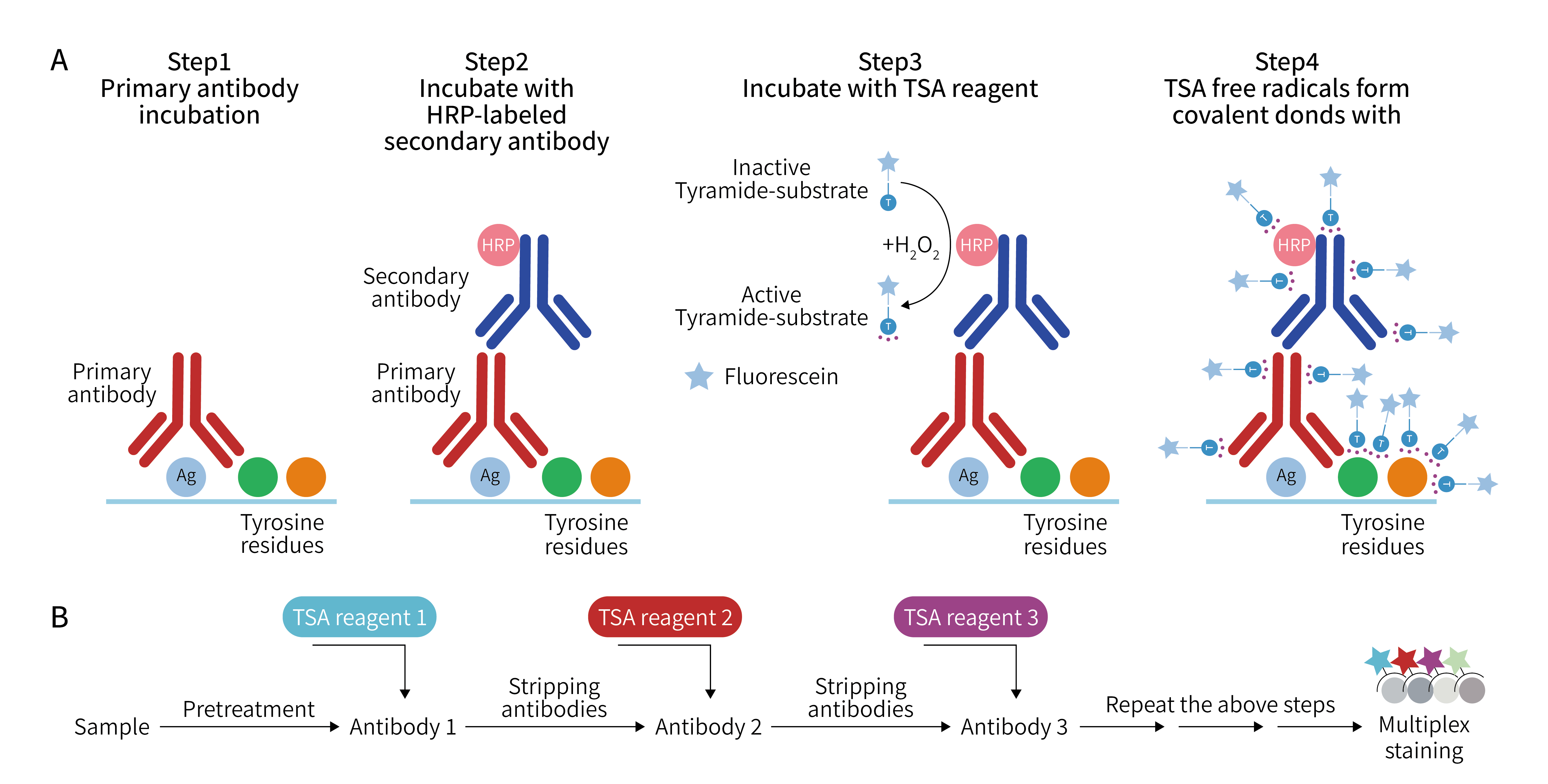
Figure 1. Schematic diagram showing the mechanism of each multiplex immunofluorescence method[1]
Brightfield Multiplex Staining IHC
To perform mIHC on FFPE tissues under brightfield microscopy, multiple chromogens/enzymes can be used. First, a specific antibody is bound to the first target in the section, and then an enzymatic reaction is used to produce a specific color at the binding site. After washing away excess antibodies, a second antibody is used to bind to the second target, producing another color. By repeating this process, different targets can be labeled with different colors under a conventional optical microscope, clearly corresponding to the morphological structure of the tissue. The greatest advantage of this approach is its intuitiveness, allowing simultaneous observation of the normal morphology of tissue cells and the distribution of targets.
Table 1. Typical chromogenic substrates in multiplex immunohistochemistry detection[2]
Substrate | Catalyst | Color |
|---|---|---|
DAB | HRP | Brown |
DAB + Ni | HRP | Black |
AEC | HRP | Red |
VIP | HRP | Purple |
NBT/BCIP | AP | Dark Blue |
Vulcan Fast Red | AP | Red |
Vector Black | AP | Black |
Nova Red | HRP | Deep Red |
TMB | HRP | Blue |
While brightfield multiplex labeling is feasible, several key factors need to be considered. The core issue is that antibodies must be conjugated with either HRP or AP to avoid cross-reactivity, which directly limits the multiplexing capability. Continuous serial IHC detection can lead to tissue degradation, further limiting the effectiveness of multiplex labeling. Additionally, there is a risk of chromogen overlap, and one color may even be masked by another, especially when chromogen spectra overlap, greatly restricting the application of brightfield chromogenic multiplex labeling. Therefore, the number of targets that can be stably detected is usually difficult to exceed 3, and there is no signal amplification effect. For extremely low-abundance targets, the color may be too faint to be visible.
Fluorescence Multiplex Labeling
To overcome the limitations of brightfield staining, fluorescence multiplex staining emerged. Instead of using colored dyes, antibodies are conjugated with fluorophores. The fluorophores for different targets have different wavelengths, and under a fluorescence microscope, these groups emit light individually. Even when 5-6 targets are labeled simultaneously, their positions can be clearly distinguished, making it particularly suitable for observing the spatial relationships between multiple targets.
However, fluorescence staining also has drawbacks. On one hand, the signal intensity of fluorescent molecules themselves is limited, and the fluorescence signal of low-abundance targets is still too weak. On the other hand, fluorescence signals of different wavelengths may bleed through, for example, the halo of red light covering the green light area, leading to misjudgment. More critically, fluorescent molecules are prone to photobleaching, requiring rapid recording during observation, otherwise the signal will become increasingly weak.
The advent of Tyramide Signal Amplification (TSA) technology has provided an efficient signal amplification solution for fluorescence multiplex staining, effectively addressing many limitations faced by traditional technologies. Its core principle is that the primary antibody specifically binds to the target, and after the secondary antibody conjugated with horseradish peroxidase (HRP) binds to the primary antibody, tyramide molecules carrying fluorophores are introduced into the reaction system. Fluorescently labeled tyramide salts produce covalent binding sites under the condition of HRP-catalyzed hydrogen peroxide, thereby triggering a large number of enzymatic reactions. These reaction products can bind to surrounding proteins (such as proteins containing tryptophan, histidine, and tyrosine residues), accumulating a large amount of fluorophores at the antigen-antibody binding site, increasing the signal intensity by tens of times, allowing even low-abundance targets to be accurately detected.
One of the significant advantages of TSA technology is its high specificity. The polymerization reaction of tyramide molecules is strictly limited to the target area where HRP is located and does not diffuse to other unrelated areas, fundamentally eliminating the phenomenon of signal cross-interference. At the same time, the fluorescence signal generated by TSA labeling has excellent stability and strong anti-quenching ability, providing good conditions for subsequent observation and imaging work. In addition, this technology has good compatibility with various fluorescent dyes, which can further expand the upper limit of the number of targets for fluorescence staining, allowing up to 8-10 targets to be labeled simultaneously, enabling comprehensive detection of multiple targets in pathological sections. In the field of scientific research, TSA technology helps researchers clearly analyze the interaction mechanisms between tumor cells and immune cells. In clinical diagnosis, it can assist doctors in more accurately identifying tumor subtypes, providing reliable experimental basis for the formulation of individualized treatment plans.
From brightfield multiplex staining to TSA-enabled multiplex fluorescence staining, the core pursuit of multiplex IHC technology has always been to "see targets more clearly, sensitively, and accurately." Today, TSA has become an indispensable key technology in fluorescence multiplex staining. It not only greatly improves the sensitivity and accuracy of target detection but also expands the application scenarios of multiplex IHC technology—from tumor research to autoimmune disease diagnosis, from basic scientific research to clinical translation, it is helping us "see through" the biological truth behind tissue sections, providing stronger support for disease diagnosis and treatment.
EnkiLife not only provides customers with a complete set of TSA multiplex labeling kits but also offers a variety of TSA specialty technical services, including IF fluorescence staining, fluorescence panoramic scanning, super multi-marker staining, and pathological analysis (5 markers and below).
For details, please check TSA mIHC Kit
References
1. Sheng W, Zhang C, Mohiuddin TM, Al-Rawe M, Zeppernick F, Falcone FH, Meinhold-Heerlein I, Hussain AF. Multiplex Immunofluorescence: A Powerful Tool in Cancer Immunotherapy. Int J Mol Sci. 2023 Feb 4;24(4):3086. doi: 10.3390/ijms24043086. PMID: 36834500; PMCID: PMC9959383.
2. Stack EC, Wang C, Roman KA, Hoyt CC. Multiplexed immunohistochemistry, imaging, and quantitation: a review, with an assessment of Tyramide signal amplification, multispectral imaging and multiplex analysis. Methods. 2014 Nov;70(1):46-58. doi: 10.1016/j.ymeth.2014.08.016. Epub 2014 Sep 19. PMID: 25242720.