A Comprehensive Guide to TSA Technology
1. Introduction to TSA Technology
Tyramide Signal Amplification (TSA) technology is a high-sensitivity in situ detection technique based on enzymatic catalytic reactions. Its core mechanism utilizes the highly efficient catalytic activity of horseradish peroxidase (HRP) to specifically and densely label target proteins or nucleic acid molecules in samples. As a key method to break through the sensitivity bottleneck of traditional detection technologies, TSA technology, through its unique signal amplification effect, can significantly enhance the intensity of target-related fluorescent signals in multiple core application scenarios such as immunocytochemistry (ICC), immunohistochemistry (IHC), and in situ hybridization (FISH). Compared with traditional detection methods, this technology can effectively capture low-abundance targets — including functionally expressed proteins, rare cell surface markers, low-copy nucleic acid fragments, and other target molecules that are difficult to detect with traditional methods, providing more precise and comprehensive detection solutions for life science research and clinical diagnosis, widely applicable to various sample types such as paraffin sections, frozen tissues, cell samples, and organoids.
2. Detection Principle of TSA Technology
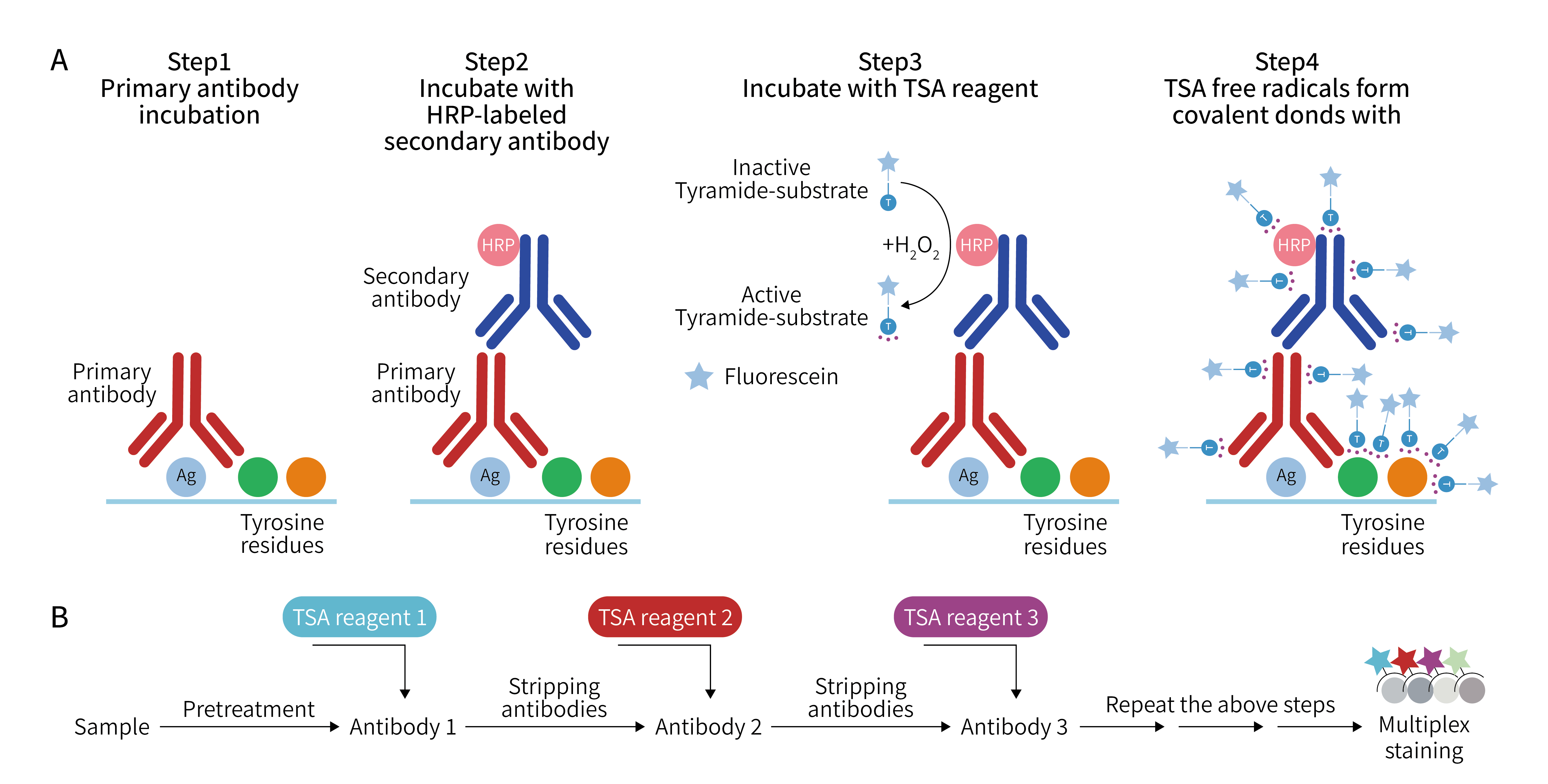
TSA technology utilizes tyramide salt to form covalent binding sites under the catalysis of HRP with H2O2, generating a large number of enzymatic products. These products can bind to surrounding protein residues (including tryptophan, histidine, and tyrosine residues), resulting in the deposition of a large amount of fluorophores at the antigen-antibody binding sites, which enhances the detection signal by 10-100 times. In simple terms, it uses HRP conjugated to the secondary antibody to catalyze the inactive fluorophores added to the system. The fluorophores are activated under the action of HRP and hydrogen peroxide, and covalently coupled to tyrosine residues of adjacent proteins, enabling stable binding between the protein sample and the fluorophores. After washing away the non-covalently bound primary antibody-secondary antibody-HRP complex, the process is repeated with another primary antibody-secondary antibody-HRP for the second round of incubation, using another tyramide fluorophore substrate, and so on to achieve multiple labeling. Since each system only contains a single antibody incubation, there is no need to worry about antibody cross-reactions or species matching issues between primary and secondary antibodies, greatly reducing the restrictions on antibody selection from different species in experimental design. That is to say, with TSA technology, all targets on the same slide can use highly specific rabbit monoclonal antibodies. Paired with the same anti-rabbit HRP secondary antibody, experiments can be conducted with significantly enhanced signal amplification.
3. Core Advantages of TSA Technology
TSA technology achieves signal amplification based on the enzymatic polymerization reaction of tyramide molecules. Its unique technical principle endows it with core competitiveness far exceeding traditional technologies, which has become a key reason for scientific research and clinical workers to choose it:
l Outstanding Signal Amplification Ability, Precisely Capturing Low-Abundance Targets
The core breakthrough of TSA technology lies in its "enzymatic signal amplification" mechanism. When HRP (horseradish peroxidase)-labeled secondary antibodies bind to primary antibodies, tyramide molecules carrying fluorophores are added, and HRP catalyzes the rapid polymerization of tyramide molecules, which stably bind to proteins around the target. This process transforms "single fluorophore labeling" into "multiple fluorophore polymerization labeling", enhancing signal intensity by tens or even hundreds of times.
This powerful amplification capability enables TSA technology to clearly detect low-abundance targets that traditional IHC cannot capture, whether they are signaling pathway proteins expressed in trace amounts within cells or specific antigens on the surface of rare immune cells, all can be precisely "illuminated", providing key evidence for early disease diagnosis and potential therapeutic target screening.
l Enhanced Multiple Labeling Capability, Enabling Panoramic Analysis of Multiple Targets
The high specificity of TSA technology is the core of its breakthrough in multiple labeling limitations. The polymerization reaction of tyramide molecules is strictly limited to the target area where HRP is located and does not diffuse to other unrelated areas, fundamentally avoiding signal cross-interference. At the same time, TSA is compatible with various fluorophores of different wavelengths, and combined with its signal amplification advantages, it can achieve synchronous labeling of 8-10 targets, far exceeding the multiple labeling limit of traditional technologies.
For example, in tumor immune microenvironment research, TSA technology can simultaneously label multiple markers such as CD3 (T cells), CD8 (cytotoxic T cells), PD-L1 (immune checkpoint molecule), CK (tumor cells), and VEGF (angiogenesis factor), clearly presenting multi-dimensional information such as immune cell infiltration patterns, interaction positions between tumor cells and immune cells, and PD-L1 expression localization, providing "panoramic" data for analyzing tumor immune escape mechanisms, which is difficult to achieve with traditional technologies.
l Gentle Operation Protects Samples, Enhancing Result Stability and Sample Utilization
Enkilife's TSA kit comes with a mild antibody elution solution that eliminates the need for high-temperature microwave treatment. It can remove unbound antibodies while completely preserving the target antigen and labeled fluorescent signals. This gentle elution method can achieve 5-7 rounds of cyclic staining, and the tissue morphology remains intact after each round of cycling, greatly reducing the risk of sample damage.
For precious samples, this advantage of TSA technology is particularly important. Only one slice is needed to complete the synchronous detection of multiple targets, eliminating the need to prepare a large number of slices and maximizing sample value. At the same time, it avoids batch errors caused by multi-slice staining, making the expression data of multiple targets based on the same tissue background, which is more comparable and the results are more reliable. The supporting fluorophore has high stability, is not easy to quench, does not require strict protection from light during experimental operation, and the stained slides can be stored at low temperature for several months.
l Wide Adaptability and Scene Compatibility, Lowering the Threshold for Technical Application
TSA technology is not limited to specific samples or detection scenarios but has strong versatility. In terms of sample types, it can be adapted to paraffin sections, frozen sections, cell slides, cell smears, tissue chips, organoids and other biological samples. In terms of detection mode, it is compatible with common equipment such as fluorescence microscopes and confocal microscopes, without the need to purchase additional specialized instruments.
In addition, TSA technology breaks the species limitation of traditional multiple staining. Enkilife's TSA kit is also equipped with secondary antibodies, breaking the traditional species limitation and being compatible with rabbit, mouse and multi-species primary antibodies, further expanding the flexibility of experimental design. Whether it is mechanism exploration in basic scientific research or precise typing in clinical diagnosis, TSA technology can quickly adapt to needs and lower the threshold for technical application.
4. Application Fields of TSA Technology
The advantages of TSA technology are not just theoretical. Its excellent performance in practical applications further confirms the necessity of choosing it:
l Applications in Scientific Research
a) Molecular Mechanism Research: Including DNA damage and repair pathway analysis, protein interactions, and detection of low-abundance proteins in signaling pathways, helping to precisely capture dynamic changes and regulatory relationships of biomolecules.
b) Cellular and Histological Analysis: Through immunofluorescence (IF) and immunohistochemistry (IHC), it enables cell subset identification, low-abundance marker localization, and spatial distribution research of neurotransmitters and receptors, providing clear visualization data for neuroscience, developmental biology and other fields.
c) Nucleic Acid Detection and Localization: Combining in situ hybridization (ISH/FISH) to enhance signals of low-copy RNA, non-coding RNA, and abnormal DNA fragments, supporting molecular genetics research such as gene expression regulation and chromosome abnormal mechanisms.
d) Cutting-edge Omics Research: As the core technical support for spatial transcriptomics and spatial proteomics, it achieves high-resolution spatial localization of genes and proteins, assisting in tissue microenvironment analysis, tumor heterogeneity research, and organ functional zoning mapping.
e) Cell Sorting and Analysis: Amplifying low-abundance cell surface marker signals in flow cytometry to precisely distinguish rare cell subsets, supporting immune cell typing, stem cell research, etc.
l Clinical Applications
a) Pathological Diagnosis and Classification: Through multiplex IHC staining, simultaneously detect multiple tumor markers and immune cell markers in clinical samples such as paraffin sections and frozen sections, assisting in tumor subtype identification, pathological grading, and prognosis assessment.
b) Early Disease Screening: Develop high-sensitivity detection platforms based on biosensing technology to capture trace abnormal biomarkers in early stages of diseases (such as tumor markers and autoantibodies), providing a basis for early warning of cancer, autoimmune diseases, etc.
c) Treatment Plan Guidance: By analyzing immune checkpoint molecules, immune cell infiltration characteristics in tumor immune microenvironment, or detecting the expression levels of drug action targets, it provides precise data for the selection and efficacy evaluation of targeted therapy and immunotherapy plans.
d) Trace Sample Detection: Suitable for precious/trace samples such as clinical puncture samples, pleural effusion cells, bone marrow smears, etc., it enables simultaneous multi-target detection while reducing sample loss, improving the utilization rate and cost-effectiveness of clinical samples.
e) Molecular Abnormality Detection: Combining FISH technology to enhance signals of low-abundance disease-causing genes and chromosome abnormal fragments, assisting in clinical diagnosis of genetic diseases and tumor-related gene mutations.
5. TSA Analysis Methods
Multiplex IHC cleverly combines four core advantages: qualitative analysis, localization, quantification, and interaction analysis. Leveraging TSA technology's efficient signal amplification effect and specific labeling capabilities, and relying on various analysis software such as HALO, Oncotopix, QuPath and other mainstream platforms, it can accurately identify the types and expression status of target antigens in tissue samples, clearly determine their specific distribution locations at cellular and subcellular levels, obtain precise values of antigen expression through standardized quantitative analysis, and deeply explore the spatial proximity relationships and functional interaction patterns between different antigens. This allows researchers and clinicians to obtain multi-dimensional, three-dimensional biomolecular information on a single section, providing comprehensive and in-depth technical support for disease mechanism research, diagnostic marker screening, and treatment effect evaluation.
a) HALO (Indica Labs, Albuquerque, New Mexico, USA, https://www.indicalab.com/halo/) is an image analysis platform used for quantitative tissue analysis in digital pathology. This software supports image alignment, segmentation, measurement, quantitative analysis, heat map analysis, cell counting and protein expression analysis, as well as cell sorting and filtering functions. HALO also comes with numerous add-on components, such as tissue classifier for automatic tissue segmentation, FISH-IF quantification for contextualizing protein and gene expression profiles for each cell, spatial analysis for identifying relative spatial distribution of cells, tissue microarray add-on for batch analysis of whole slide TMA images, and serial section analysis for analyzing stained serial sections to obtain additional IHC markers. These add-on components are also compatible with various image and digital slide formats, namely JPG, TIF, ND2, MRXS, QPTIFF, component TIFF, VSI, NDPI, NDPIS, SVS, AFI, CZI, SCN, LIF and BIF. More importantly, HALO itself is compatible with Vectra and Insituplex and is often used as a recommended imaging platform.
b) Oncotopix (Visiopharm, Hoersholm, Denmark; https://www.visiopharm.com/) is a commercially available quantitative digital pathology image analysis software. By integrating image analysis, machine learning and artificial intelligence technologies, it can efficiently complete the analysis, management and report generation of massive datasets, and seamlessly connect with laboratory information management systems, compatible with all image/slide formats. Compared with similar software, the significant advantage of this software is that it has obtained in vitro diagnostic (IVD) certification and is used as a compliant device in various clinical detection scenarios such as breast cancer and lung cancer. Oncotopix's "Invasive Tumor Detection APP" can realize automated and precise detection of invasive tumors, integrate complete biomarker analysis process, automatically identify lesion hotspots, quantify biomarker expression levels, and visually present tumor heterogeneity.
c) HistoCAT is a freely accessible interactive computing platform capable of quantitative analysis of highly multiplexed, single-cell resolution tissue samples. This platform integrates high-dimensional image visualization technology, cell phenotype characteristic analysis methods, and innovative algorithms for comprehensively studying cell-cell interactions and cell social networks in complex tissues. In addition, HistoCAT is equipped with an innovative algorithm that can detect neighboring cell interactions that occur more frequently than expected by random probability, and identify more significant interaction relationships and unique cellular microenvironments in the entire dataset and cohort. However, HistoCAT has limitations in tissue segmentation function and needs to be used in conjunction with other cell segmentation software such as CellProfiler.
d) QuPath (https://qupath.github.io) is another high-throughput free image analysis software suitable for whole slide image analysis, with powerful batch processing and script writing capabilities. This software can process large-scale whole slide imaging data and conduct subsequent analysis without adjusting the image data to a processable size through cropping or downsampling. However, this software has certain limitations in digitally scoring biomarkers with complex staining patterns (such as mismatch repair proteins, immune checkpoint inhibitors like PD-L1). Because the staining of such biomarkers involves multiple cell populations including tumor cells and immune cells, the mottled staining distribution pattern significantly increases the difficulty of distinguishing staining signals between the two types of cells.
Table 1 Some Commonly Used Multiplex IHC/IF Analysis Platforms
Characteristic | Analysis software | |||
|---|---|---|---|---|
HALO | Oncotopix | HistoCAT | QuPath | |
Open source | No | No | Yes | Yes |
Advantages | Recommended software for Vectra and InSituPlex. Widely adopted and compatible with most multiplex IHC/IF platforms. | Widely adopted and compatible with most multiplex IHC/IF platforms. | Recommended software for IMC | Supports most image formats and platforms, such as ImageJ, MATLAB, CellProfiler, etc. Widely adopted and compatible with most multiplex IHC/IF platforms. Capable of processing whole slide imaging data. Built-in cell segmentation software. |
Limitations | Paid | Paid | Needs to be used in conjunction with other segmentation software like CellProfiler | Code/programming based |
Developer | Indica Labs | Visionpharm | Bodenmiller Lab | P. Bankhead and team |
EnkiLife not only provides customers with a complete set of TSA multiplex labeling kits but also offers a variety of TSA specialty technical services, including IF fluorescence staining, fluorescence panoramic scanning, super multi-marker staining, and pathological analysis (5 markers and below).
Product | Catalog Number |
|---|---|
TSA Six-Label Seven-Color Multiplex Immunohistochemistry Kit | |
TSA Five-Label Six-Color Multiplex Immunohistochemistry Kit | |
TSA Four-Label Five-Color Multiplex Immunohistochemistry Kit | |
TSA Three-Label Four-Color Multiplex Immunohistochemistry Kit | |
TSA Two-Label Three-Color Multiplex Immunohistochemistry Kit |
For details, please check TSA mIHC Kit