Aurora kinases
About Aurora Kinases
Aurora kinases belong to a highly conserved family of mitotic serine/threonine kinases with three members identified among mammals: Aurora A, B, and C. Studies on the temporal expression pattern and subcellular localization of Aurora kinases in mitotic cells suggest an association with mitotic structure.
Aurora kinase functional influences span from G2 phase to cytokinesis and may be involved in key cell cycle events such as centrosome duplication, chromosome bi-orientation and segregation, cleavage furrow positioning, and ingression.
Aurora A is detected at the centrosomes, along mitotic spindle microtubules, and in the cytoplasm of mitotically proliferating cells. Aurora A protein levels are low during G1 and S phases and peak during the G2/M phase of the cell cycle. Phosphorylation of Aurora A at Thr288 in its catalytic domain increases kinase activity. Aurora A is involved in centrosome separation, maturation, and spindle assembly and stability.
Expression of Aurora B protein also peaks during the G2/M phase of the cell cycle; Aurora B kinase activity peaks at the transition from metaphase to the end of mitosis. Aurora B associates with chromosomes during prophase prior to relocalizing to the spindle at anaphase. Aurora B regulates chromosome segregation through the control of microtubule-kinetochore attachment and cytokinesis.
Expression of both Aurora A and Aurora B during the G2/M phase transition is tightly coordinated with histone H3 phosphorylation; research investigators have observed overexpression of these kinases in a variety of human cancers.
Aurora C localizes to the centrosome from anaphase to cytokinesis and both mRNA and protein levels peak during G2/M phase. Although typical Aurora C expression is limited to the testis, research studies report overexpression of Aurora C is detected in various cancer cell lines.
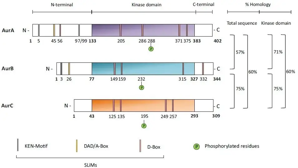
Structure Domain Composition of Aurora Kinases A-C
Relevant Antibodies
| Catalog# | Product Name | Reactivity | Application |
|---|---|---|---|
| APRab04258 | ARK-1 (phospho Thr288) Rabbit Polyclonal Antibody | Human,Mouse,Rat | WB,IHC-P,IF-P,IF-F,ICC/IF,ELISA |
| AMRe21613 | Aurora A Rabbit Monoclonal antibody | Human | WB,IHC,IF,IP,ELISA |
| AMRe21394 | Aurora B Rabbit Monoclonal antibody | Human | WB,IHC,IF,IP,ELISA |
| APS0635 | HRP-conjugated Polyclonal Goat Anti-Rabbit IgG(H+L) Secondary Antibody | ELISA, WB, Dot blotRabbit | ELISA, WB, Dot blot |
| AMre80004 | GAPDH (12R9) Rabbit Monoclonal Antibody | WB,ELISA Human,Mouse,Rat,Rabbit,Dog,Monkey | WB,ELISA |
| APRab04258 | ARK-1 (phospho Thr288) Rabbit Polyclonal Antibody | Human,Mouse,Rat | WB,IHC-P,IF-P,IF-F,ICC/IF,ELISA |
Related Products
References
- Warner SL, Bearss DJ, Han H, Von Hoff DD. Targeting Aurora-2 kinase in cancer. Mol Cancer Ther. 2003 Jun;2(6):589-95. [PMID: 12813139].
- Katayama H, Brinkley WR, Sen S. The Aurora kinases: role in cell transformation and tumorigenesis. Cancer Metastasis Rev. 2003 Dec;22(4):451-64. [PMID: 12884918].
- Andrews PD, Knatko E, Moore WJ, Swedlow JR. Mitotic mechanics: the auroras come into view. Curr Opin Cell Biol. 2003 Dec;15(6):672-83. [PMID: 14644191].
- Pascreau G, Arlot-Bonnemains Y, Prigent C. Phosphorylation of histone and histone-like proteins by aurora kinases during mitosis. Prog Cell Cycle Res. 2003;5:369-74. [PMID: 14593731].
- Crosio C, Fimia GM, Loury R, et. Mitotic phosphorylation of histone H3: spatio-temporal regulation by mammalian Aurora kinases. Mol Cell Biol. 2002 Feb;22(3):874-85. [PMID: 11784863]
- Kimura M, Matsuda Y, Yoshioka T. Cell cycle-dependent expression and centrosome localization of a third human aurora/Ipl1-related protein kinase, AIK3. J Biol Chem. 1999 Mar 12;274(11):7334-40.[PMID: 10066797].
| Flora Flora is a technical support expert at EnkiLife, familiar with immunology and neuroscience, dedicated to providing customers with high-quality product combinations and technical support to help achieve research in neurodegenerative diseases and other neuroscience areas. |
