C-33A cells are a human cell line derived from cervical cancer, initially established by N. Auersperg from cervical epithelial cells of a 66 year old white patient with cervical squamous cell carcinoma. These cells initially exhibit a hypodiploid karyotype and epithelial cell morphology, but with continuous passage, the karyotype becomes unstable. C-33A cells exhibit abnormal expression of retinoblastoma protein (pRB), upregulation of p53, and point mutations at codon 273. These cells are insensitive to human papillomavirus DNA and RNA, but tested negative for mycoplasma.
C-33A cells have the following biological characteristics:
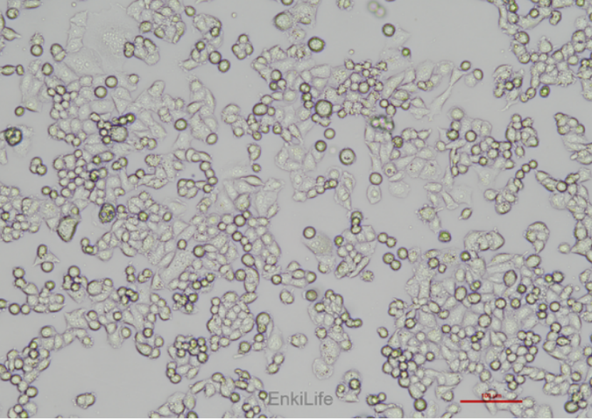
1. Morphological features: adherent growth, presenting as epithelioid polygonal cells, low-density growth presenting as epithelioid polygonal cells, high-density growth presenting as spherical and adherent cells.
2. Genetic features: Abnormal expression of pRB, upregulation of p53, and presence of a point mutation at codon 273. [1]
3. Karyotype characteristics: Initially, it was a hypodiploid karyotype, but with passage, the karyotype became unstable.
4. Tumor formation ability: Undifferentiated cancer can be formed in nude mice, with lymphatic and blood metastasis ability, and has been used for the first time to establish a metastasis model. [2]
5. Drug sensitivity: Shows different survival advantages for certain drugs such as ER, LA, and AST. [7]
C-33A cells are widely used in cancer research, especially in the study of biological processes such as cell proliferation, migration, invasion, and apoptosis. [4,5,6]
The Application Fields of C-33A Cells in Medical Research
The application areas of C-33A cells in medical research mainly include the following aspects:
1. Research on tumor metastasis and spread:
OC-33A cells have been used to study the metastatic ability of cervical cancer cells, particularly the mechanisms of lymphatic and blood borne metastasis. Research has shown that C-33A cells, after subcutaneous implantation in nude mice, are studied through pathways such as subcutaneous tumor formation, lymphatic and blood metastasis. [2]
By genetic modification, such as integrating the mRFP1 gene into C-33A cells, real-time monitoring of tumor growth and metastasis can be achieved. [2]
2. Research on virus therapy:
OC-33A cells were used to evaluate the efficacy of viral therapy. For example, using attenuated recombinant cowpox virus GLV-1h68 to infect C-33A cells, it was found that the virus has therapeutic potential for primary tumors of various cancers, especially in eliminating metastasis in the C-33A model. [2]
3. Drug sensitivity and resistance research:
Research has found that baicalin can increase the sensitivity of cisplatin resistant cervical cancer C-33A cells to cisplatin by promoting intracellular autophagy levels to enhance their sensitivity to cisplatin.
The combination of propofol and chemotherapy drug paclitaxel can significantly inhibit the proliferation and induce apoptosis of C-33A cells, reversing drug resistance. [17]
4. Research on gene expression and signaling pathways:
Research has found that overexpression of non receptor tyrosine kinase BMX in C-33A cells can promote their migration and invasion abilities. [9]
The high expression of omiR-155 in C-33A cells is associated with cell proliferation, promotion of epithelial mesenchymal transition (EMT), and inhibition of apoptosis. [12]
OGC1qR plays a key role in HPV16 E6 and E7 gene mediated apoptosis of C-33A cells, with mitochondrial dysfunction being its main mechanism. [3]
5. Molecular mechanism research:
Research has found that AMP bidirectionally regulates autophagy and apoptosis in C-33A cells through the PI3K/Akt/mTOR pathway, revealing its potential therapeutic targets in cervical cancer. [10]
OmiR-3681 negatively regulates the proliferation and migration of C-33A cells by inhibiting the PI3K/Akt pathway. [15]
6. Cell culture and basic research:
OC-33A cells are an adherent cervical cancer cell line widely used in laboratory scientific research, including cell culture, gene transfection, drug screening, and more.
In summary, C-33A cells have important application value in multiple fields such as tumor metastasis, viral therapy, drug sensitivity, gene expression, and signaling pathways.
Latest research progress on C-33A cells
Based on the latest research progress of C-33A cells, the following are some key findings:
1. AMP regulates autophagy and apoptosis in C-33A cells through the PI3K/Akt/mTOR pathway:
Research has shown that AMP can significantly inhibit the growth of C-33A cells and induce apoptosis of C-33A cells through death receptors or mitochondrial pathways. AMP induced autophagy occurs through the PI3K/Akt/mTOR pathway, and inhibition of the PI3K/Akt/mTOR pathway enhances AMP induced autophagy, while its activation enhances AMP's autophagy inhibition. [10]
2. The effect of traditional Chinese medicine ingredients on C-33A cells:
Research has shown that baicalin can increase the LC3-II/LC3I ratio of cisplatin resistant cervical cancer C-33A cells, promote the expression of Beclin-1, ATG5, and ATG12 in cells, and thus enhance cellular autophagy levels. [8]
3. Transfer ability of C-33A cells:
Research has shown that C-33A cells exhibit significant lymphatic and blood metastasis ability after subcutaneous implantation of tumor cells in nude mice. The study also found that attenuated recombinant cowpox virus GLV-1h68 can effectively inhibit the metastasis of C-33A cells in tumor models. [2]
4. Effects of MIR22HG on C-33A cells:
Research has shown that overexpression of MIR22HG can significantly inhibit the proliferation and migration ability of C-33A cells.
5. The role of gC1qR in C-33A cells:
Research has found that gC1qR expression decreases in C-33A cells transfected with HPV-16, and the accumulated gC1qR protein can induce cell apoptosis. Mitochondrial functional protection can reverse gC1qR induced apoptosis in C-33A cells. [3]
6. Effects of propofol on C-33A cells:
Research has shown that propofol can significantly reduce the viability of C-33A cells and increase cell apoptosis. In addition, propofol can also inhibit the expression of HOTAIR protein. [14]
7. The effect of FBXO31 on the proliferation of C-33A cells:
Research has found that FBXO31 can inhibit the proliferation of C-33A cells, and the inhibition rate is concentration - and time-dependent. FBXO31 is mainly expressed in the nucleus and is less expressed in cervical cancer tissues than in adjacent normal tissues. [13]
8. The role of circMYLK in C-33A cells:
Research has shown that circMYLK accelerates the development of cervical cancer by upregulating RHEB and activating the mTOR signaling pathway. The expression of circMYLK is high in C-33A cells, and its transcription and translation levels are both high. [16]
These studies provide important scientific basis for understanding the biological characteristics and potential therapeutic targets of C-33A cells
References:
1.The state of the p53 and retinoblastoma genes in human cervical carcinoma cell lines. [PMID: 1648218]
2. Characterization of metastasis formation and virotherapy in the human C33A cervical cancer model. [PMID: 24887184]
3. The role of gC1qR in regulating survival of human papillomavirus 16 oncogene-transfected cervical cancer cells. [PMID: 21725590]
4. Long Non-Coding RNA TMPO-AS1 Promotes Cervical Cancer Cell Proliferation, Migration, and Invasion by Regulating miR-143-3p/ZEB1 Axis. [PMID: 32184662]
5. MicroRNA-378 enhances migration and invasion in cervical cancer by directly targeting autophagy-related protein 12. [PMID: 29488616]
6. Increase of IFN-γ and TNF-α production in CD107a + NK-92 cells co-cultured with cervical cancer cell lines pre-treated with the HO-1 inhibitor. [PMID: 25302050]
7. HER Family Receptors are Important Theranostic Biomarkers for Cervical Cancer: Blocking Glucose Metabolism Enhances the Therapeutic Effect of HER Inhibitors. [PMID: 28255362]
8. Research progress on the regulation of autophagy in tumor cells by Chinese herbal medicine. He Man, Xu Haibo et al. Chinese Herbal Medicine 2021, Vol.52, No.10, pp.3142-3150.
9. Li Yuanyuan, Kou Jiushe, Wu Tao et al. Non-receptor tyrosine kinase Etk/BMX promotes the migration and invasion of cervical cancer cells [J]. Chinese Journal of Metastatic Tumors, 2019, 02(4) : 31-35.
10. PI3K/Akt/mTOR-mediated bidirectional regulation ofAmpelopsin from Nekemias megalophylla modulates autophagy and apoptosis in cervical cancer. Shiyi Xu et al. [2024-11-26]
11. Effect of hyperoside on cervical cancer cells and transcriptome analysis of differentially expressed genes.[ PMID: 31516392]
12. MiR-155 Promotes Proliferation and Epithelial-mesenchymal Transition (EMT) and Inhibits Apoptosis of Cervical Cancer Cells via Regulating ING4. Min Du et al.Int J Hum Genet, 22(1): 1-7 (2022). IJHG.
13. Study on the effect of FBXO31 on the proliferation of human cervical cancer cells C-33A and its expression and clinical significance in human cervical cancer tissues. Qin Xiaoyang et al. International Journal of Obstetrics and Gynecology.
14. Jin Baowei, Wang Kai, Guo Jianrong et al. Advances in the study of the effects of propofol on tumor growth and metastasis mechanisms [J]. Chinese Journal of Clinical Pharmacology and Therapeutics, 2024, 29(9): 1042-1048.
15. Retracted Article: RNA-sequencing identified miR-3681 as a negative regulator in the proliferation and migration of cervical cancer cells via the posttranscriptional suppression of HGFR. [PMID: 35519460]
16. circRNA MYLK Accelerates Cervical Cancer via Up-Regulation of RHEB and Activation of mTOR Signaling. [PMID: 32547198]
17. Protamine sulfide enhances ferroptosis in paclitaxel-induced cervical cancer cells by inhibiting GPX4 expression. Zhao Mengyun et al. Department of Anesthesiology, Peking Union Medical College Hospital, Chinese Academy of Medical Sciences.
18. Global cancer statistics 2018: GLOBOCAN estimates of incidence and mortality worldwide for 36. cancers in 185 countries.