Frozen Sample Immunohistochemistry (IHC) Detection
1. Tissue OCT Embedding
Purpose: Embed tissue samples with OCT compound and freeze for subsequent sectioning and fixation.
Materials:
- Tissue samples
- Isopentane
- Dry ice
- OCT compound
- Embedding molds
Procedure:
Prepare a metal container, partially fill with isopentane and place on dry ice, cool for 5 minutes.
Place fresh tissue into embedding mold and fill with OCT compound, adjusting tissue orientation.
Position the mold above the cooled isopentane container for 10-20 seconds until the tissue block becomes opaque, avoiding direct contact between isopentane and OCT.
Store frozen tissue blocks at -80°C freezer for future use, or at -20°C for short-term storage.
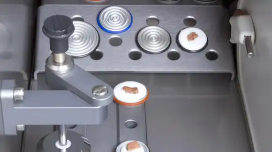
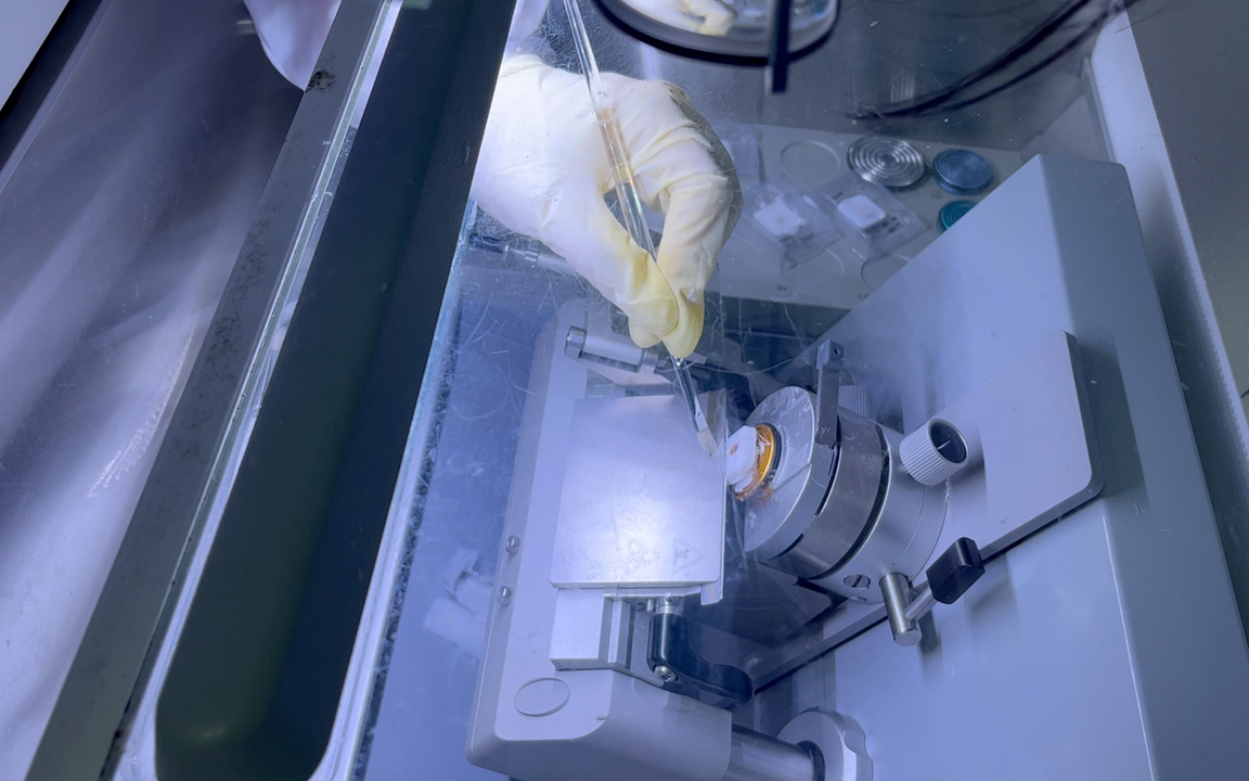
2. Sectioning
Purpose: Cut embedded tissue into thin sections and mount on slides.
Materials:
- OCT-embedded tissue samples
- Microscope slides
- Cryostat and blades
Procedure:
Place OCT-embedded tissue in cryostat chamber at -20°C for temperature equilibration (30min-1h). For larger or special tissue types, extend to 2h if needed.
Secure frozen tissue block on specimen holder.
Adjust cryostat blade, ensure proper fixation and set appropriate cutting angle.
Trim tissue block to expose tissue surface, typically 10-30μm thickness.
Cut sections at 5-20μm thickness (adjust according to experiment), discard initial potentially problematic sections.
Transfer sections to slides using a brush, prepare for subsequent fixation steps.
3. Fixation
Purpose: Preserve tissue morphology through fixative treatment.
Materials:
- Tissue sections
- Buffer (PBS)
- Fixatives (10% neutral buffered formalin, 4% paraformaldehyde, 100% acetone or methanol)
Procedure:
Allow freshly cut frozen sections to sit at room temperature for 15 minutes.
Select appropriate fixative based on detection target:
For low molecular weight proteins, peptides and enzymes: 10% neutral buffered formalin or 4% paraformaldehyde
For high molecular weight protein antigens: 100% acetone or methanol
For high molecular weight protein antigens: 100% acetone or methanol
Immerse tissue sections in fixative, incubate at room temperature for 15 minutes.
Wash sections three times with PBS, 3min each.
4. Blocking
Purpose: Reduce background staining through blocking steps.
Materials:
- Tissue sections
- Blocking solution (e.g. 2-10% normal serum)
- Wash buffer (PBS)
Procedure:
Wash slides three times with PBS, 3min each.
Block endogenous peroxidase activity by incubating with 3% hydrogen peroxide for 10 minutes, then wash with PBS.
Perform protein blocking by incubating with normal serum blocking solution for 30 minutes.
5. Antibody Incubation
Purpose: Stain tissue with antibodies, using either directly conjugated primary antibodies or indirect secondary antibody detection.
Materials:
- Antibody dilution buffer
- Wash buffer (PBS)
- Conjugated primary antibody
Procedure:
Determine optimal antibody dilution according to manufacturer's instructions and dilute in antibody dilution buffer.
Apply diluted primary antibody to tissue, incubate at room temperature for 1 hour or at 4°C overnight.
Wash sections three times with PBS, 3min each.
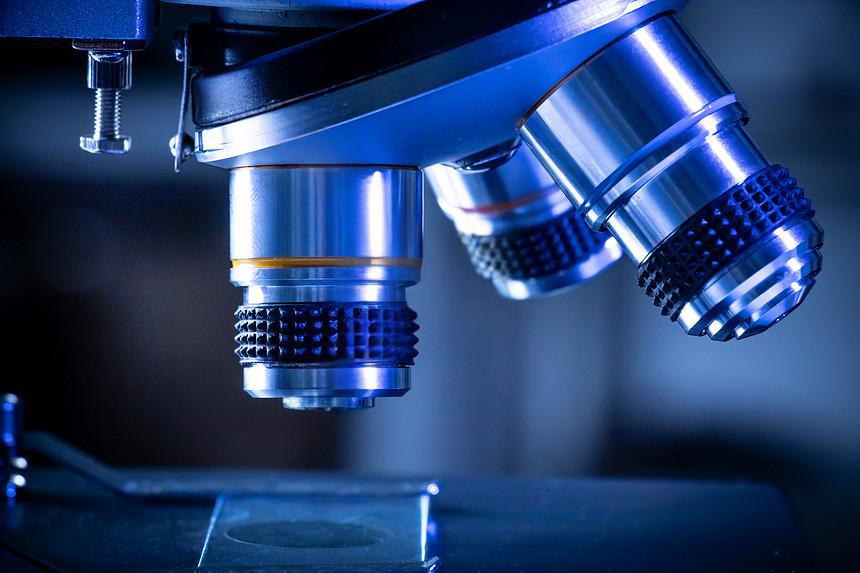
6. Chromogenic Reaction & Microscopy
Purpose: Visualize staining results through chromogenic reaction.
Materials:
- Chromogenic substrate (e.g. DAB, AEC)
- Counterstain (optional, e.g. hematoxylin)
- Mounting medium (optional, e.g. aqueous mounting medium)
- Coverslips
- Microscope
Procedure:
Apply prepared chromogen solution to tissue sections, monitor under microscope until desired positive signal appears.
Wash sections three times with PBS, 3min each.
Apply counterstain solution to sections, incubate at room temperature for 2-5 minutes.
Rinse slides with PBS to remove excess stain and blue hematoxylin.
Apply small amount of mounting medium to slides.
Carefully place coverslips over sections using forceps, avoiding air bubbles.
Examine sections under microscope, or store at 4°C if not immediately observed.
 | Jeff Jeff is a pathology product expert at EnkiLife, specializing in immunohistochemistry research and product development for over 12 years. He has a high level of insight into user needs and is committed to helping our clients conduct immunohistochemistry experiments and accelerate scientific discoveries. |