Cell Fluorescent Dyes
Cell Fluorescent Dyes are detection tools developed based on the principles of fluorescent chemistry, the characteristics of biomolecules, and the structural features of cells. They are specifically designed for labeling and observing various structures and molecules within cells. These dyes are widely used in cellular morphology, cell function analysis, cell signaling, live imaging, and multiplex labeling.
EnkiLife Cell Fluorescent Dyes, with their outstanding performance and diverse options, provide strong support for life science research.
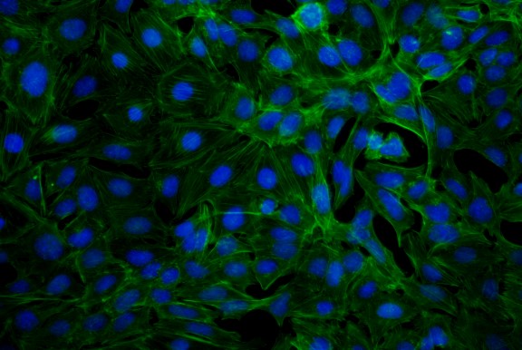
Cytoskeleton Dyes

Cytoskeleton fluorescent dyes are essential tools for labeling and visualizing the cytoskeleton structures within cells. They play a significant role in various fields such as cell morphology, cell function analysis, cell signaling, live imaging, and multiplex labeling. The cytoskeleton is composed of actin, tubulin, and intermediate filaments. These structures are crucial for maintaining cell shape, cell motility, cell division, and signal transduction.
| Cat# | Product Name | Excitation Wavelength | Emission Wavelength |
|---|---|---|---|
| RA20015 | Rhodamine-Phalloidin | 546nm | 575nm |
| RA20011 | Fluorescein 555-Phalloidin | 555nm | 565nm |
| RA20010 | Fluorescein 488-Phalloidin | 490nm | 515nm |
| RA20014 | Fluorescein 680-Phalloidin | 681nm | 698nm |
| RA20013 | Fluorescein 633-Phalloidin | 630nm | 650nm |
| RA20012 | Fluorescein 594-Phalloidin | 590nm | 617nm |
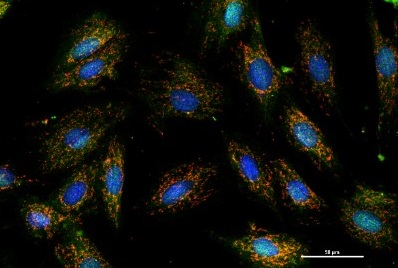
Subcellular Structure Dyes

Subcellular structure fluorescent dyes are fluorescent markers used to label and observe various organelles and structures within cells. They are of significant importance to cell biology research. These dyes provide powerful tools for cell biologists, enabling the visualization of different structures and functions within cells through appropriate selection and application.
| Cat# | Product Name | Excitation Wavelength | Emission Wavelength |
|---|---|---|---|
| RA20020 | Mitochondrial Tracker (Red) | 579nm | 599nm |
| RA20021 | Mitochondrial Tracker (Green) | 490nm | 523nm |
| RA20022 | JC-1 (Mitochondrial Membrane Potential Probe) | 550nm / 485nm | 600nm / 535nm |
| RA20024 | DASPEI (Mitochondrial Fluorescent Probe) | 461nm | 589nm |
| RA20025 | 4-Di-1-ASP (Mitochondrial Fluorescent Probe) | 474nm | 606nm |
| RA20026 | Lysosome Tracker (DeepRed) | 649nm | 665nm |
| RA20027 | Lysosome Tracker (Red) | 577nm | 590nm |
| RA20028 | Lysosome Tracker (Green) | 504nm | 511nm |
| RA20030 | Dihydroethidium (Hydroethidine;DHE) | 518nm | 610nm |
| RA20031 | Fluo-3 AM (2mM), Calcium Ion Fluorescent Probe | 506nm | 526nm |
| RA20032 | Fluo-4 AM (2mM), Calcium Ion Fluorescent Probe | 494nm | 516nm |
| RA20033 | Fluo-4 AM, Calcium Ion Fluorescent Probe | 494nm | 516nm |
| RA20034 | Fluo-3 AM, Calcium Ion Fluorescent Probe | 518nm | 610nm |
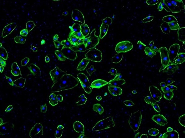
Cell Membrane Dyes

Cell membrane fluorescent dyes are fluorescent markers used to label cell membranes and other lipophilic biological structures. Common cell membrane fluorescent dyes include DiD, DiR, DiO, and DiI dyes, which are a group of lipophilic fluorescent dyes. Once these dyes enter the cell membrane, they laterally diffuse across the entire membrane, and at optimal concentrations, they can stain the entire cell membrane. These dyes come in different fluorescence colors, making them suitable for multicolor fluorescence imaging analysis and flow cytometry in live cells. They provide great convenience for the study of cell membrane morphology and structure.
| Cat# | Product Name | Excitation Wavelength | Emission Wavelength |
|---|---|---|---|
| RA20019 | Enhanced DiO Membrane Probe (Green) | 484nm | 501nm |
| RA20001 | DiA Membrane Probe (Green) | 456nm | 590nm |
| RA20002 | Dilinoleyl DiI Membrane Probe (Orange-Red) | 549nm | 565nm |
| RA20003 | DiD Membrane Probe (Red) | 644nm | 663nm |
| RA20004 | DiI Membrane Probe (Orange-Red) | 549nm | 565nm |
| RA20005 | DiO Membrane Probe (Green) | 484nm | 501nm |
| RA20006 | DiR Membrane Probe (Near-Infrared) | 748nm | 780nm |
| RA20007 | CM-DiI Membrane Probe (Orange-Red) | 549nm | 565nm |
| RA20008 | CM-DiD Membrane Probe (Red) | 644nm | 663nm |
| RA20009 | CM-DiD Membrane Probe (Orange-Red) | 549nm | 565nm |
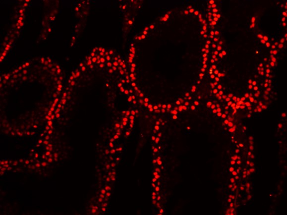
Nuclear Dyes

Nuclear fluorescent dyes are a class of dyes that can bind to DNA in the cell nucleus and emit fluorescence. They play an important role in cell biology research. Commonly used nuclear fluorescent dyes include DAPI, Hoechst 33258, Hoechst 33342, PI (Propidium Iodide), and others. These nuclear fluorescent dyes provide a variety of options for observing and studying the cell nucleus due to their distinct fluorescence and staining properties.
| Cat# | Product Name | Excitation Wavelength | Emission Wavelength |
|---|---|---|---|
| RA20035 | DAPI Nuclear Staining Solution (Ready-to-use) | 360nm | 460nm |
| RA20036 | DAPI Nuclear Dye | 360nm | 460nm |
| RA20037 | Hoechst 33342 Nuclear Staining Solution (Ready-to-use) | 346nm / 350nm | 460nm / 461nm |
| RA20038 | Hoechst 33258 Nuclear Staining Solution (Ready-to-use) | 346nm / 352nm | 460nm / 461nm |
| RA20039 | Hoechst 33342 Live Cell DNA Dye | 346nm / 350nm | 460nm / 461nm |
| RA20040 | Hoechst 33258 Live Cell DNA Dye | 346nm / 352nm | 460nm / 461nm |
| RA20041 | Propidium Iodide (PI) | 493nm / 535nm | 636nm / 617nm |
| RA20042 | dsDNA Quantification Reagent (Green) | 480nm | 520nm |
| RA20067 | DRAQ5 Live Cell DNA Staining Solution | 647nm | 681nm |
| RA20068 | High Sensitivity dsDNA Quantification Kit | 485nm | 530nm |
| RA20016 | 5(6)-CFDA SE Live Cell Fluorescent Probe | 490-500nm | 517-520nm |
| RA20017 | 6-CDCFDA SE Live Cell Fluorescent Probe | 504nm | 529nm |
Product Advantages
- Specific Binding: Cell fluorescent dyes can specifically bind to target cell structures or molecules, such as cell membranes, nuclei, mitochondria, lysosomes, etc. This ensures that the signal originates only from the target area, with minimal background fluorescence.
- High Sensitivity: The intensity of the fluorescent signal is proportional to the concentration or quantity of the target molecules. Even at low concentrations, the signal can be detected by high-sensitivity microscopes or imaging devices.
- Low Cytotoxicity: Optimized chemical design ensures minimal impact on cellular physiological functions, making the dyes suitable for long-term live-cell imaging.
- High Stability: The fluorescent signal remains stable under various experimental conditions, such as different pH levels, temperatures, and light exposure, with minimal quenching.
- Wide Wavelength Range: Cell fluorescent dyes cover a broad range of wavelengths from visible to near-infrared light (400-750 nm), meeting the needs of both single-color and multiplex labeling experiments.
- Low Background Fluorescence: Near-infrared fluorescent dyes (e.g., 690-750 nm) exhibit lower background fluorescence and higher tissue penetration capabilities, making them ideal for live imaging.
- Easy to Use: Most cell fluorescent dyes have simple application methods with minimal staining steps and no complex pre-treatment required.
- Strong Compatibility: These dyes are compatible with common cell fixatives, permeabilization agents, and other fluorescent markers, allowing seamless integration into various experimental workflows.
