Basic information ofHuman ovarian cancer cell Skov3
Detailed origin and historical background of SKOV3 cell line:
The SKOV3 cell line is a human ovarian cancer cell line, originally isolated by G. Trempe and L. J. Old in 1973 from ascites samples of ovarian adenocarcinoma in a 64 year old white female patient. This cell line has an epithelial cell like morphology and exhibits characteristics of adherent growth under culture conditions.
The establishment of SKOV3 cell line was carried out at a time when the different histological types of ovarian cancer were not fully understood. Therefore, many cell lines used for ovarian cancer research were isolated from patient samples at earlier time points. Nevertheless, the SKOV3 cell line has been extensively studied for its similarity to ovarian cancer, including its abnormal cell growth and differentiation, anti apoptotic ability, and ability to invade surrounding tissues.
In addition, the SKOV3 cell line exhibits resistance to various cytotoxic drugs, such as tumor necrosis factor, diphtheria toxin, cisplatin, and doxorubicin. These characteristics make the SKOV3 cell line an important tool for studying drug resistance and treatment strategies in ovarian cancer.
The detailed origin and historical background of the SKOV3 cell line can be traced back to 1973, when G. Trempe and L. J. Old isolated it from ascites samples of ovarian adenocarcinoma in a 64 year old white female patient.
The SKOV3 cell line has significant value in ovarian cancer research due to its unique biological characteristics and wide range of applications.

Cultivation conditions:
The temperature of the incubator should be strictly controlled at 37℃, with temperature fluctuations not exceeding ± 0.5℃, simulating the physiological environment of the human body. Carbon dioxide (CO₂) concentration: 5%, maintaining the pH stability of the culture medium (usually pH between 7.2-7.4). Generally, a normoxic environment (21% O₂) is used, and special experiments (such as hypoxic models) can be adjusted to low oxygen (such as 1-5% O₂). The relative humidity inside the incubator should be kept above 95% to avoid evaporation of the culture medium, which may cause an increase in osmotic pressure. RPMI-1640 or DMEM/F12 (1:1 mixture) are commonly used as culture media, both of which can support their growth (different laboratories may have slight differences due to passage history, it is recommended to refer to the recommended information of cell sources).
Adding 10% fetal bovine serum (FBS) requires selecting qualified serum that has undergone heat inactivation (56℃, 30 minutes) to remove complement and other components, reducing stimulation to cells. To prevent pollution, penicillin (100 U/mL) and streptomycin (100 μg/mL) can be added. It is recommended to regularly replace antibiotic free culture media for long-term cultivation to avoid drug resistance.
Application in medicine:
SKOV3 cells have been used in various experiments in research, including drug sensitivity testing, signaling pathway studies, and exploration of tumor development mechanisms. For example, studies have shown that ginsenoside Rh3 can inhibit the proliferation of SKOV3 cells by initiating the apoptotic pathway [15]; Pinecone chrysanthemum glycoside reduces the survival rate of SKOV3 cells by inhibiting cell proliferation and stem cell like properties [10].

In addition, arsenic trioxide can inhibit the proliferation and metastasis of SKOV3 cells by downregulating the expression of VEGF gene.
SKOV3 cells also exhibit resistance to certain drugs. For example, SKOV3 cells can induce resistance to cisplatin through moderate dose and intermittent action methods [14]. In addition, BMSCs (bone marrow mesenchymal stem cells) can promote the proliferation, migration, and invasion ability of SKOV3 cells through various mechanisms.
In terms of signaling pathways, there are multiple important signaling pathways in SKOV3 cells, such as JAK2/STAT3 [17], PI3K/AKT [6], Wnt/β - catenin [3,13], etc. Activation or inhibition of these pathways may affect cell proliferation, apoptosis, and migration ability. For example, ESC-3 induces apoptosis in SKOV3 cells through the Wnt/β - catenin and Notch signaling pathways [3].
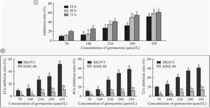
The effect of Jimatone regulating the JAK2/STAT3 signaling pathway on the proliferation, migration, and invasion of ovarian cancer SKOV3 cells.
What are the specific applications and results of SKOV3 cell line in drug sensitivity testing?
The specific application and results of SKOV3 cell line in drug sensitivity testing are as follows:
1. Cisplatin sensitivity:
OSKOV3 cells exhibit high sensitivity to cisplatin (DDP). Research has shown that SKOV3 cells are significantly inhibited by cisplatin at temperatures below 41℃, with a sensitivity increase of 79.885% [25]
However, when SKOV3 cells develop into a drug-resistant cell line (SKOV3/DDP), their sensitivity to cisplatin significantly decreases. The resistance index of SKOV3/DDP cells is 2.21, indicating strong resistance to cisplatin [24].
The impact of MFG-E8 gene silencing:
The silencing of MFG-E8 gene can affect the sensitivity of SKOV3 cells to cisplatin. The experimental results showed that after silencing the MFG-E8 gene, SKOV3 cells showed increased sensitivity to cisplatin [23].
3. Reversal of resistance by rapamycin:
Rapamycin can reverse the resistance of SKOV3/DDP cell lines to cisplatin. At a concentration of 25 μg/L of rapamycin, the inhibition rate of SKOV3/DDP cells against cisplatin significantly increased, reaching 81.43%. In addition, rapamycin promotes cell apoptosis and reverses drug resistance by inhibiting the expression of p-mTOR and p-p70s6k proteins in the Akt/mTOR pathway [24].
4. Multidrug resistance:
OSKOV3 cells also exhibit sensitivity to other anticancer drugs such as paclitaxel and doxorubicin. However, the SKOV3 multidrug-resistant cell line (SKOV3 ts) established by the concentration gradient increasing method showed higher resistance to these drugs [27].
5. Endoplasmic reticulum stress and cisplatin resistance:
Under the action of cisplatin, the expression of endoplasmic reticulum stress markers Grp78 and PDI in oSKOV3 cells significantly increased, and the expression of apoptosis signaling molecules such as CHOP and Caspase-4 also increased, indicating that the endoplasmic reticulum stress pathway plays an important role in cisplatin sensitivity.
6. The effect of hyperthermia combined with cisplatin:
The combination of hyperthermia and cisplatin can significantly increase the sensitivity of SKOV3 cells to cisplatin and reverse the drug resistance of SKOV3/DDP cells. The optimal experimental conditions are 41 ℃ and 90 minutes of thermal therapy time. [25]
In summary, the SKOV3 cell line exhibits sensitivity to multiple anticancer drugs in drug sensitivity testing, but its sensitivity to certain drugs significantly decreases as resistance develops.
How to induce resistance of SKOV3 cells to cisplatin through experimental methods?
The resistance of SKOV3 cells to cisplatin can be induced through the following experimental methods:
1. Repeat cross subcutaneous injection of human ovarian cancer SKOV3 cells labeled with green fluorescent protein (GFP), and transplant tumor fragments into nude mice. Through this method, two SKOV3/DDP I and SKOV3/DDP II cell lines can be established, which exhibit significant resistance to cisplatin [29].
2. Treat SKOV3 cells with different concentrations of cisplatin. For example, SKOV3 cells can be treated with cisplatin concentrations of 1, 2, 4, 8, 16, 32, 64, and 128 μg/ml for 24 hours, and then their half maximal inhibitory concentration (IC50) can be measured. The results showed that with the increase of cisplatin concentration, the IC50 value of SKOV3 cells gradually increased, indicating that the cell's resistance to cisplatin gradually increased [30].
3. Co culture with tumor associated fibroblasts (CAFs). Research has shown that CAFs can promote the proliferation and migration of SKOV3 cells, and reduce their sensitivity to cisplatin. Specifically, the IC50 value of SKOV3 cells co cultured with CAFs was significantly higher than that of SKOV3 cells cultured alone [31].
4. Conduct gene overexpression experiments. For example, SKOV3 cell lines overexpressing RRM2 gene can be constructed by lentiviral transfection and evaluated for their sensitivity to cisplatin. The results showed that SKOV3 cells overexpressing the RRM2 gene exhibited significantly reduced sensitivity to cisplatin [34].
5. Changes in endoplasmic reticulum stress (ERS) functional status. Research has shown that cisplatin can induce ERS in SKOV3 cells, and the combination of ERS inducer TG and cisplatin treatment can increase the expression of ERS related proteins and apoptosis rate in SKOV3 cells [38].
6. Regulating mitochondrial function through the HSP90/SIRT3/SOD2 pathway. Research has found that the sensitivity of SKOV3 cells to the HSP90 inhibitor SNX2112 is similar to that of cisplatin, and mitochondrial function and protein input ability may be the main reasons for SKOV3 cells' resistance to cisplatin [37].
What is the specific mechanism of action of JAK2/STAT3, PI3K/AKT, Wnt/β - catenin signaling pathways in SKOV3 cell line?
The specific mechanisms of action of JAK2/STAT3, PI3K/AKT, and Wnt/β - catenin signaling pathways in SKOV3 cell line are as follows:
1. JAK2/STAT3 signaling pathway:
Activation mechanism: When cytokines (such as IL-6) bind to receptors on the cell surface, they cause receptor dimerization and recruit related JAKs (Janus kinases). Subsequently, activation of JAK leads to receptor tyrosine phosphorylation, forming binding sites for STAT. STATs (signal transduction and transcription activators) are phosphorylated by tyrosine, dissociate from receptors, enter the nucleus, bind to DNA, and regulate gene transcription.
Function and impact: The JAK2/STAT3 signaling pathway plays an important role in cell proliferation, migration, and invasion. For example, melatonin can inhibit the epithelial mesenchymal transition (EMT) of ovarian cancer SKOV3 cells induced by IL-6 by suppressing the JAK2/STAT3 pathway, reducing the number of migrating cells, Vimentin protein levels, and the expression of p-JAK2/JAK2 and p-STAT3/STAT3, while increasing E-cadherin protein levels [41]. In addition, Germacrone can also inhibit the proliferation, migration, and invasion of SKOV3 cells by downregulating the activity of the JAK2/STAT3 signaling pathway [39].
2. PI3K/AKT signaling pathway:
Activation mechanism: PI3K (phosphatidylinositol 3-kinase) converts phosphatidylinositol into PIP3 (phosphatidylinositol-3,4,5-triphosphate), activating protein kinase B (Akt). Akt is fully activated after phosphorylation on the cell membrane, and subsequently phosphorylates and activates various proteins such as FOXO, Bad, etc., regulating cellular function.
Function and impact: The PI3K/AKT signaling pathway plays a critical role in cell survival, proliferation, and metabolism. For example, FGF3 and FGF10 regulate the migration and invasion ability of SKOV3 cells through the Wnt/β - catenin signaling pathway [42]. In addition, there is a cross regulatory relationship between the PI3K/AKT signaling pathway and the JAK2/STAT3 signaling pathway. The JAK2/STAT3 signaling pathway can activate the PI3K/AKT signaling pathway, thereby providing upstream initiating factors [43].
3. Wnt/β - catenin signaling pathway:
Activation mechanism: Wnt protein binds to Frizzled receptors on the cell surface, activating signaling molecules such as Disheveled (Dvl) and subsequently activating β - catenin. β - catenin accumulates inside the cell and enters the nucleus, activating downstream gene expression.
Function and impact: The Wnt/β - catenin signaling pathway plays an important role in determining cell fate, proliferation, and cancer development. For example, FGF3 and FGF10 regulate the migration and invasion ability of SKOV3 cells through the Wnt/β - catenin signaling pathway [42]. In addition, Akt inhibits the activity of GSK-3 β (a negative regulator of β - catenin) by phosphorylating it, causing β - catenin to accumulate in the cell and enter the nucleus, activating downstream gene expression.
In summary, the JAK2/STAT3, PI3K/AKT, and Wnt/β - catenin signaling pathways regulate cell proliferation, migration, invasion, and EMT processes in the SKOV3 cell line through different mechanisms.
How do BMSCs promote the proliferation, migration, and invasion ability of SKOV3 cells?
Based on the provided information, it is not possible to directly answer how BMSCs promote the proliferation, migration, and invasion ability of SKOV3 cells. However, we can extract some possible mechanisms and impacts from relevant research.
1. Inhibit the proliferation and apoptosis of SKOV3 cells:
A study has shown that BM MSCs (bone marrow mesenchymal stem cells) can induce apoptosis in SKOV3 cells. Specifically, after co culturing BM MSCs with SKOV3 cells, flow cytometry analysis revealed a significant increase in the apoptosis rate of SKOV3 cells [44]. This suggests that BMSCs may inhibit the proliferation of SKOV3 cells through some mechanism.
2. The effect of mechanical stretching on BMSCs:
Another study explored the effect of mechanical stretching on BMSCs and found that mechanical stretching inhibits the invasive ability of BMSCs by downregulating MT1-MMP through the PI3K/Akt signaling pathway [45]. Although this study mainly focuses on the effect of mechanical stretching on BMSCs, it can be speculated that similar signaling pathways may also affect the behavior of BMSCs under other conditions.
3. The effect of tumor associated cells on SKOV3 cells:
A study based on a tumor microenvironment model showed that tumor associated cells can significantly enhance the migration and invasion ability of SKOV3 cells [47]. Although this study did not directly involve BMSCs, it provides an important background that other cell types in the tumor microenvironment can significantly affect the behavior of SKOV3 cells.
Although there is no direct evidence to suggest how BMSCs promote the proliferation, migration, and invasion ability of SKOV3 cells, it can be speculated that BMSCs may affect the behavior of SKOV3 cells by regulating specific signaling pathways or interacting with other cells in the tumor microenvironment.
References
1. Bone Marrow Mesenchymal Stem Cells Promote Ovarian Cancer Cell Proliferation via Cytokine Interactions. [PMID: 38928452]
2. Cyclin D1 expression and the inhibitory effect of celecoxib on ovarian tumor growth in vivo. [PMID: 21152316]
3. ESC-3 induces apoptosis of human ovarian carcinomas through Wnt/β-catenin and Notch signaling in vitro and in vivo.[PMID: 27878242]
4. FOXO3a-mediated suppression of the self-renewal capacity of sphere-forming cells derived from the ovarian cancer SKOV3 cell line by 7-difluoromethoxyl-5,4'-di-n-octyl genistein. [PMID: 24604613]
5. Anti-tumorigenic and Platinum-Sensitizing Effects of Apolipoprotein A1 and Apolipoprotein A1 Mimetic Peptides in Ovarian Cancer. [PMID: 30745873]
6. Effect of oleanol on the proliferation, migration, invasion and apoptosis of human ovarian cancer SKOV3 cells. Dai Linghong et al.[2019]
7. Melittin-MIL-2 fusion protein as a candidate for cancer immunotherapy. [PMID: 27246873]
8. MiR-144-3p targets SGK3 and inhibits the growth and invasion of ovarian cancer through the Hippo signaling pathway. Yao Hairong et al.[2019-12-31]
9. SRC-3/TRAF4 facilitates ovarian cancer development by activating the PI3K/AKT signaling pathway.[ PMID: 36625999]
10. Limonidin inhibits the proliferation, invasion and stem cell-like characteristics of ovarian cancer SKOV3 cells. Dong Yana et al.[2021-12-31]
11. Inoculation of human ovarian cancer cell SKOV3 in BalB/c mice.[2022-09-14]
12. Chen Zhiwei, Mei Qingbu, Zhang Qi et al. Effects of Tribulus saponins on the proliferation of human ovarian cancer cells SKOV3 [J]. Chinese Journal of Gerontology,2013.
13. Study on the interaction between epidermal growth factor receptor 2 and Wnt/β-catenin signal to promote ovarian cancer cell metastasis. Liu Fenfen et al..[2018-03-15]
14. Establishment and study of in vitro drug-resistant model of ovarian carcinoma. Li Hong et al.[2011-02-28]
15. Yi Wengong, Zhu Baocheng, Xu Haitao et al. The inhibitory effect of ginsenoside Rh3 on the proliferation of human ovarian cancer cell SKOV-3 [J]. Chinese Journal of Maternal and Child Health,2018.
16. Tumorigenesis and metastasis inhibition of METCAM/MUC18 in human ovarian cancer SKOV3 cells.[PMID: 35299043]
17. Tang Xin, Han Fengjuan, Li Wei et al. Study on the effect of Curcuma alcohol on JAK2/STAT3 signaling pathway in human ovarian cancer SKOV3 cell line [J]. Chinese Journal of Obstetrics and Gynecology,2013.
18. Zhou Xiangyu, Liu Ning, Yan Shan et al.S1 induces apoptosis in human ovarian cancer cells via the mitochondrial pathway [J]. Chinese Journal of Gerontology,2014.
19. Shen Wei, Liang Bingfeng, Qin Jinjin et al. Effects of NaD and cisplatin on proliferation and apoptosis of SKOV3 ovarian cancer cells [J]. Chinese Journal of Gerontology,2016.
20. He Kai, Zhang Guirong, Yang Shuli et al. Effect of arsenic trioxide on VEGF gene expression in human ovarian cancer cells [J]. Chinese Journal of Laboratory Medicine,2008.
21. Establishment and validation of a novel invasion-related gene signature for predicting the prognosis of ovarian cancer. [PMID: 35292033]
22. Liu Lifeng, Zhong Luting, Zhu Weipei. Expression and mechanism of 7-dehydrocholesterol reductase in epithelial ovarian cancer cell line SKOV3 [J]. Chinese Journal of Experimental Surgery., 2022, 39(12) : 2421-2424.
23. Li Na, Dai Congwei, et al. The effect of MFG-E8 on cisplatin sensitivity in ovarian cancer SKOV3 cells and its molecular mechanism [J]. Cancer Prevention and Treatment Research,, 2021, 48(5): 464-469.
24. Xu Guocai, Wang Dongyan, Lu Huaiwu, et al. Discussion on the mechanism of rapamycin reversal of SKOV3/DDP cisplatin resistance in ovarian cancer cell lines [J]. Journal of Sun Yat-sen University (Medical Science Edition), 2018,39(1).
25. Ma Yan, Qu Xinxin, Zhang Xiuyan. In vitro study on the effect of ovarian cancer cells on cisplatin sensitivity during intraperitoneal hyperthermia chemotherapy [J]. Chinese Journal of Clinical Oncology, 2010, 37(20): 1153-1156.
26Deng Yao, Wu Jiang, Yin Guangfu. Induction culture and identification of tumor-associated cells and their effects on SKOV3 cell biology [J]. Clinical Medical Progress, 2020, 10(12): 3048-3059.
27. Yang Yan et al. Study on the targeted reversal of ovarian cancer multidrug resistance by folic acid modified chitosan small interfering RNA nanoparticles[2009-05-01]
28. Li Hong, Zhao Liyan, Qiao Xinmin. Establishment of cisplatin-resistant ovarian cancer cell lines [J]. Journal of Dalian Medical University, 2001, 23(1).
29. Establishment of SPD-resistant ovarian epithelial carcinoma cell line and its nude mouse xenograft model and discussion on drug resistance characteristics. Shi Lijun et al.
[2014-07-25]
30. Effect of apatinib combined with fluoroparaparib on proliferative capacity of cisplatin-resistant cells in human ovarian carcinoma.[2023-07-28]
31. Study on tumor-associated fibroblasts and cisplatin resistance in epithelial ovarian carcinoma. Jiang Yu et al.[2024-07-19]
32. ABT-737 enhances cisplatin sensitivity in human ovarian cancer cells and induces mitochondrial division. Journal of Toxicology.[2018-06]
33. Upregulation of p27Kip1 by demethylation can sensitize SKOV3 cells, which are resistant to cisplatin, to ovarian cancer. Yan Zhao etal.[2016-06-18]
34. Construction of SKOV3 ovarian cancer cell line with overexpression of RRM2 gene and observation on sensitivity to cisplatin.[2017]
35. Effect of genistein on proliferation, apoptosis and cisplatin sensitivity of SKOV-3 drug-resistant ovarian cancer cells.[2006-03-05]
36. Study on the mechanism of mitochondrial protein input regulated by HSP90/SIRT3/SOD2 pathway in ovarian cancer cell drug resistance.[2011]
37. Study on the mechanism of mitochondrial protein input regulated by HSP90/SIRT3/SOD2 pathway in ovarian cancer cell drug resistance[2023-09-11]
38. Effect of endoplasmic reticulum functional status on cisplatin sensitivity of ovarian cancer cells. Zhou Xiaoshui et al.[2015]
39. Effect of JAK2/STAT3 signaling pathway regulated by Germacrone on cell viability of ovarian cancer SKOV3[J]. ACADEMIC JOURNAL OF CHINESE PLA MEDICAL SCHOOL, 2021, 42(8): 866-872.
40. Melatonin attenuates IL-6-induced epithelial-to-mesenchymal transition via inhibition of JAK2/STAT3 signaling pathway in ovarian cancer SKOV3 cells.
41. Melatonin inhibits IL-6 induced epithelial-mesial transformation of SKOV3 ovarian cancer cells by inhibiting JAK2/STAT3 pathway.《Chinese Journal of Reproductive Health》2019 , (05) : 431-435.
42.Effect of FGF3 and FGF10 on cell migration and invasion of human ovarian cancer SKOV3 cells and its relationship with Wnt/β-catenin signaling pathway. Wang Jia et al43. The PI3K/Akt, p38MAPK, and JAK2/STAT3 signaling pathways mediate the protection of SO2 against acute lung injury induced by limb ischemia/reperfusion in rats.
[PMID: 26541157]
44. Anti-proliferative effects of mesenchymal stem cells (MSCs) derived from multiple sources on ovarian cancer cell lines: an in-vitro experimental study.[ PMID: 31351482]
45. MT1-MMP downregulation via the PI3K/Akt signaling pathway is required for the mechanical stretching-inhibited invasion of bone-marrow-derived mesenchymal stem cells.
[PMID: 30659604]
46. Strategic targeting of miR-183 and β-catenin to enhance BMSC stemness in age-related osteoporosis therapy. [PMID: 39277663]
47. Induction and identification of tumor-associated cells and their effects on SKOV3 cell biology.[2020-12-07]