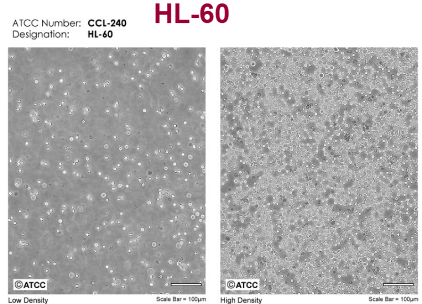
The HL-60 cell line is a human primary myeloid leukemia cell line derived from acute myeloid leukemia (AML). This cell line was initially obtained by S.J. Collins et al. from a 36 year old female patient with acute promyelocytic leukemia and has been widely used to study the differentiation, proliferation, apoptosis, and drug sensitivity of myeloid cells [9].
HL-60 cells have the ability to self differentiate and can be induced to differentiate into mature granulocytes, monocytes, and macrophages through various chemical inducers such as dimethyl sulfoxide (DMSO), all trans retinoic acid (ATRA), 1,25-dihydroxyvitamin D3, etc. [5,6,10]. These inducers promote the differentiation of HL-60 cells by activating specific signaling pathways, such as retinoic acid receptor (RAR) - mediated signaling [7,12].
HL-60 cells exhibit high heterogeneity during the culture process, with some cells differentiating into macrophage like or eosinophil like phenotypes [9]. In addition, HL-60 cells exhibit characteristics such as phagocytic activity, chemotaxis, superoxide production, and NBT dye reduction assay, making them an ideal model for studying the function of myeloid cells [6].
In drug research, HL-60 cells are widely used to evaluate the activity of anticancer drugs. For example, α - tomatine has been shown to inhibit the growth of HL-60 cells and induce their apoptosis, involving the activation of NF - κ B and its effects on apoptosis related proteins such as Bcl-2 and Survive [2]. In addition, some studies have also explored the cytotoxic effects of other compounds such as Lp16-PSP on HL-60 cells, and found that it induces cell death through the apoptotic pathway [8].
The HL-60 cell line has become an important tool for understanding the pathogenesis, drug screening, and new drug development of myeloid leukemia due to its wide application and multifunctionality in leukemia research.
The HL-60 cell line is a widely used model for studying the differentiation program of human leukemia, and its differentiation mechanism involves multiple signaling pathways and molecular mechanisms. In particular, retinoic acid (RA) - mediated signaling plays a crucial role in the differentiation of HL-60 cells.
Retinoic acid promotes the differentiation of HL-60 cells through signaling pathways mediated by its receptors (RAR α, RXR α, and RXR γ). Specifically, after binding to RAR α, RA activates downstream transcription factors such as nuclear factor kappa B (NF κ B) and transcription factor ERK, thereby regulating gene expression [17,19,20]. The activation of these transcription factors promotes the expression of key genes involved in cell cycle arrest and differentiation, such as BLR1 and CXCR5 [19,22].
RA induced signaling also involves the MAPK signaling pathway, particularly the activation of ERK. The activation of ERK not only enhances cell differentiation, but also further enhances RA induced signaling through a positive feedback mechanism [19,20]. In addition, RA can also inhibit tumor growth and angiogenesis by suppressing Rb RAF interactions [17].
In addition, the differentiation process induced by RA also involves other important signaling molecules and pathways. For example, RA can promote nuclear accumulation of Raf1 and enhance signal transduction along the c-Raf/GSK-3/VDR axis [17]. In addition, RA can prevent cell apoptosis by inhibiting the c-Raf/MEK/ERK signaling pathway [17].
In summary, retinoic acid promotes the differentiation of HL-60 cells through its receptor-mediated signaling pathway, particularly by activating transcription factors such as NF κ B and ERK, as well as regulating the MAPK signaling pathway.
How is the heterogeneity exhibited by HL-60 cells during the culture process generated, and what impact does this heterogeneity have on research?
According to the search results, the main focus is on the culture conditions, differentiation process, and some experimental results of HL-60 cells, but there is no detailed explanation of the mechanism of cell heterogeneity and its specific impact on research.
However, some possible reasons and impacts can be inferred from some evidence:
1. Differences in culture conditions: HL-60 cells exhibit different growth characteristics under different culture conditions. For example, differences in culture medium, pH value, temperature, and carbon dioxide concentration may lead to cellular heterogeneity [24]. These changes in conditions may affect the physiological state and behavior of cells, resulting in heterogeneity.
2. Changes during differentiation: HL-60 cells can differentiate by adding different stimuli (such as ATRA, DMSO, etc.), and the gene expression and phenotype of the cells will change during differentiation [23,25,26]. The changes during this differentiation process may also lead to cellular heterogeneity.
3. Differences in experimental procedures: Different experimental procedures (such as washing, centrifugation, staining, etc.) may also affect cell heterogeneity [23]. For example, different washing steps may lead to differences in cell surface markers, thereby affecting cell heterogeneity.
4. Spontaneous differentiation of cells: HL-60 cells have the characteristic of spontaneous differentiation, and even without external stimulation, cells may spontaneously differentiate into different subtypes [15]. This spontaneous differentiation is also a possible reason for heterogeneity.
5. Impact of research: Heterogeneity of cells may affect the interpretation and application of experimental results. For example, when using HL-60 cells as a model for research, heterogeneity may lead to inconsistent experimental results, affecting the reliability and reproducibility of the study [26].
In summary, although the searched information did not directly answer the question, it can be inferred that the heterogeneity of HL-60 cells may be caused by various factors, including culture conditions, differentiation processes, experimental procedures, and spontaneous differentiation of cells.
What is the specific mechanism of α - tomatine in inhibiting the growth and inducing apoptosis of HL-60 cells?
The specific mechanisms by which α - tomatine inhibits the growth and induces apoptosis of HL-60 cells involve multiple aspects, including interactions with cell membrane associated cholesterol, changes in mitochondrial membrane potential, and activation of the NF - κ B signaling pathway.
Alpha tomato alkaloids significantly inhibit the growth of HL-60 cells and induce apoptosis by interacting with cholesterol in the cell membrane. Research has shown that the apoptosis and growth inhibition of HL-60 cells treated with α - tomato alkaloids are partially blocked by the cholesterol scavenger 12-O-deacetyl-13-cholesterolide, indicating that the interaction between α - tomato alkaloids and cell membrane associated cholesterol plays an important role in regulating their effects [2].
In addition, α - lycopene induces cell apoptosis by affecting mitochondrial membrane potential. Research has found that in the presence of alpha tomato alkaloids, the mitochondrial membrane potential of HL-60 and K562 cell lines changes from a green curve to a blue curve, indicating a loss of mitochondrial membrane potential [27]. This change in mitochondrial function further supports the possibility of alpha tomato alkaloids inducing cell apoptosis through the mitochondrial pathway.
Alpha tomato alkaloids may also exert their effects by activating the NF - κ B signaling pathway. Research has shown that alpha tomato alkaloids can activate NF - κ B, thereby promoting cell apoptosis [2]. This mechanism may be related to the regulation of cell membrane cholesterol by alpha tomato alkaloids, as cholesterol regulation can affect the activation of NF - κ B [2].
Although the induction of apoptosis in HL-60 cells by α - tomato alkaloids is not related to caspase activation, its induction of apoptosis in other types of leukemia cells such as MCF-7 and K562 is related to caspase activation [27,28]. This indicates that the mechanism of action of α - tomato alkaloids may differ in different cell types.
Alpha tomato alkaloids significantly inhibit the growth and induce apoptosis of HL-60 cells through various mechanisms such as interacting with cell membrane cholesterol, affecting mitochondrial membrane potential, and activating the NF - κ B signaling pathway.
What is the specific mechanism of the cytotoxic effect of Lp16-PSP on HL-60 cells and its induction of apoptosis pathway?
The specific mechanism of the cytotoxic effect of Lp16-PSP on HL-60 cells and its induction of apoptosis pathway is as follows:
1. Cytotoxic effects:
Lp16 PSP exhibits significant cytotoxicity in HL-60 cells. Research has shown that Lp16 PSP can cause significant changes in the morphology and volume of HL-60 cells, including a decrease in cell volume and bleeding, indicating that the cells are undergoing apoptosis or death processes [8].
2. Mechanisms of inducing apoptosis:
Lp16 PSP induces apoptosis in HL-60 cells through various pathways:
ODNA fragmentation: After treatment with Lp16 PSP, the DNA of HL-60 cells undergoes fragmentation, which is an important feature of apoptosis [8].
Mitochondrial membrane potential loss: Lp16 PSP leads to mitochondrial membrane potential loss, which is the result of the combined action of exogenous and endogenous apoptotic pathways [8].
Ocaspase activation: Lp16 PSP activates caspase-8 and caspase-9, further promoting the activation of caspase-3, which is a key step in cell apoptosis [8].
Cell cycle arrest: Lp16 PSP mediates G1 phase cell cycle arrest through p21WAF1/CIP1, thereby inhibiting cell proliferation and promoting apoptosis [8].
Inhibition of NF - κ B: Lp16 PSP inhibits the translocation of NF - κ B to the nucleus by reducing the level of phosphorylated I κ B α, thereby reducing the expression of genes related to apoptosis and proliferation, such as Bcl-2 and Cyclin D1 [8].
3. Changes in gene expression:
After treatment with Lp16 PSP, significant changes were observed in the expression of genes related to apoptosis and cell cycle. Specifically, it manifests as upregulation of Bax, caspase-3, caspase-8, caspase-9, FasL, p21, p27, and Bcl-2, while downregulation of Bcl-2, cdk4, cdk6, cdk2, cyclin D1, and E [8].
What are the latest developments in the application of HL-60 cell line in drug development?
The application of HL-60 cell line in new drug development mainly focuses on the following aspects:
1. Drug screening and biological activity evaluation:
The HL-60 cell line is widely used for drug screening and biological activity evaluation. For example, researchers tested the biological activity of the novel anti-cancer drug iPAdo using the HL-60 cell line [5]. In addition, the HL-60 cell line has also been used to evaluate the effects of novel oxaphosphoramide drugs (such as D-18864, D-17272, and D-19575) on cell survival rate, cell size, and cell cycle [33].
2. Immunological research:
The HL-60 cell line can induce the differentiation of mature granulocytes, monocytes, macrophages, and eosinophils, and can be used to study immunological processes such as cell migration and chemical signaling [5]. In addition, the HL-60 cell line has been used to study Toll like receptors (TLRs) and cytokine expression, such as the regulatory effect of baicalin on TLRs and cytokine expression [29].
3. Study on the mechanism of apoptosis:
The HL-60 cell line is an ideal model for studying cell apoptosis. Research has shown that the HL-60 cell line responds to various apoptotic stimuli and can be used to study the positive and negative regulatory mechanisms of exogenous or physiological pharmacological stimuli on apoptosis [16]. In addition, the HL-60 cell line can also be used to study the effect of inflammatory agents on apoptosis regulation [16].
4. Gene expression and differentiation research:
The HL-60 cell line has bidirectional differentiation potential and can be induced into monocytes or granulocytes, making it an ideal model for studying myeloid cell differentiation [32]. The HL-60 cell line has also been used to study the heterogeneity of antibacterial activity, functional response, and gene expression [30].
5. Other applications:
The HL-60 cell line can also be used to study the cryopreservation and recovery functions of granulocytes [30], as well as to investigate cell proliferation, differentiation, and apoptosis events [31].
References
1. Croton caudatus Geiseler Induces Differentiation of the Acute Myeloid Leukemia cell lines. Takhellambam Chanu Machathoibi et al.
2. α-Tomatine inhibits growth and induces apoptosis in HL-60 human myeloid leukemia cells. HUARONG HUANG et al. [PMID: 25625536]
3. Nuclear Proteomics of Induced Leukemia Cell Differentiation. Svetlana Novikova et al. [PMID: 36291090]
4. Antileukemic activity of combined epigenetic agents, DNMT inhibitors zebularine and RG108 with HDAC inhibitors, against promyelocytic leukemia HL-60 cells. [PMID: 22820861]
5. N6-isopentenyl adenosine,a promising anticancer agent:synthesis of novel analogues,
Evaluation of antiproliferative activity and insight into action mechanism. Università degli Studi di Milano et al.[2008-12-31]
6. Choosing the Right Cell Line for Acute Myeloid Leukemia (AML) Research. Rafał Skopek et al.[PMID: 36982453]
7. (Chalcogen)semicarbazones and their cobalt complexes differentiate HL-60 myeloid leukaemia cells and are cytotoxic towards tumor cell lines. [PMID: 30108695]
8. Lp16-PSP, a Member of YjgF/YER057c/UK114 Protein Family Induces Apoptosis and p21WAF1/CIP1 Mediated G1 Cell Cycle Arrest in Human Acute Promyelocytic Leukemia (APL) HL-60 Cells. Thomson Patrick Joseph et al.
9. HOXA9 as a Risk Factor in Acute Myeloid Leukaemia. Chris Spearman.
10. Molecular and epigenetic alterations in normal and malignant myelopoiesis in human leukemia 60 (HL60) promyelocytic cell line model. Somnath Paul et al. [PMID: 36819104]
11. Cyclooxygenase-2 (COX-2) inhibition constrains indoleamine 2,3-dioxygenase 1 (IDO1) activity in acute myeloid leukaemia cells.[ PMID: 23973990]
12.An Effective Model of the Retinoic Acid Induced HL-60 Differentiation Program. Ryan Tasseff et al. [PMID: 29085021]
13. The effect of bone marrow mesenchymal stromal cell exosomes on acute myeloid leukemia's biological functions: a focus on the potential role of LncRNAs. [PMID: 38777995]
14. Intracellular Accumulation and Secretion of YKL-40 (CHI3L1) in the Course of DMSO-Induced HL-60 Cell Differentiation. [PMID: 38675403]
15. Human acute promyelocytic leukemia cell HL-60 specification. Chinese Academy of Sciences Committee for the Preservation of Typical Culture.
16. In vitro detection of apoptotic stimuli by use of the HL-60 myeloid leukemic cell line.[PMID: 8548540]
17. Roscovitine enhances all- trans retinoic acid (ATRA)-induced nuclear enrichment of an ensemble of activated signaling molecules and augments ATRA-induced myeloid cell differentiation.[PMID: 32256976]
18. Retinoic acid receptor regulation of decision-making for cell differentiation. Mustapha Najimi et al. [PMID: 37082619]
19. Nuclear Raf-1 kinase regulates the CXCR5 promoter by associating with NFATc3 to drive retinoic acid-induced leukemic cell differentiation. [PMID: 24330068]
20. An Effective Model of the Retinoic Acid Induced HL-60 Differentiation Program. [PMID: 29085021]
21. Human myeloblastic leukemia cells (HL-60) express a membrane receptor for estrogen that signals and modulates retinoic acid-induced cell differentiation. [PMID: 18692045]
22. Modeling and analysis of retinoic acid induced differentiation of uncommitted precursor cells. [PMID: 21437295]
23. Neutrophil extracellular traps-mediated Beclin-1 suppression aggravates atherosclerosis by inhibiting macrophage autophagy. [PMID: 35923856]
24.Model regulatory networks for proteins that are activated and inhibited in the process of induced granulocyte differentiation. S.E. Novikova et al.
25. Combined Effect of Naturally-Derived Biofilm Inhibitors and Differentiated HL-60 Cells in the Prevention of Staphylococcus aureus Biofilm Formation. [PMID: 33182261]
26. Transcriptomic analysis of lipoteichoic acid‑treated undifferentiated and neutrophil‑like differentiated HL‑60 cells. [PMID: 38476893]
27. α-Tomatine-mediated anti-cancer activity in vitro and in vivo through cell cycle- and caspase-independent pathways. [PMID: 22970166]
28. The cytotoxic effect of α-tomatine in MCF-7 human adenocarcinoma breast cancer cells depends on its interaction with cholesterol in incubation media and does not involve apoptosis induction. [PMID: 24100733]
29. Andrographolide modulate some toll-like receptors and cytokines expressions in HL-60 cell line. Ajit Kumar Thakur et al.Pharm Pharmacol Int J. 2015;2(4):116‒120.
30. What Is the Evolutionary Fingerprint in Neutrophil Granulocytes?. Leonie Fingerhut et al.[PMID: 32630520]
31. Involvement of intrinsic mitochondrial pathway in neosergeolide-induced apoptosis of human HL-60 leukemia cells: the role of mitochondrial permeability transition pore and DNA damage. [PMID: 22775415]
32. Effects of extracts from regenerating organisms on differentiation of the human myeloid leukaemia HL-60 cell line.
33.In vitro effects of new generation oxazaphosphorines on human promyelocytic leukemia cells. [PMID: 23767290]